

Holter monitor (24h)
Ambulatory electrocardiography; Electrocardiography - ambulatory; Atrial fibrillation - Holter; Flutter - Holter; Tachycardia - Holter; Abnormal heart rhythm - Holter; Arrythmia - Holter; Syncope - Holter; Arrhythmia - HolterA Holter monitor is a machine that continuously records the heart's rhythms. The monitor is worn for 24 to 48 hours during normal activity.
How the Test is Performed
Electrodes (small conducting patches) are stuck onto your chest. These are attached by wires to a small recording monitor. You carry the Holter monitor in a pocket or pouch worn around your neck or waist. The monitor runs on batteries.
While you wear the monitor, it records your heart's electrical activity.
- Keep a diary of what activities you do while wearing the monitor, and how you feel.
- After 24 to 48 hours, you will return the monitor to your health care provider's office.
- Your provider will look at the records and see if there have been any abnormal heart rhythms.
It is very important that you accurately record your symptoms and activities so the provider can match them with your Holter monitor findings.
Electrodes must be firmly attached to the chest so the machine gets an accurate recording of the heart's activity.
While wearing the device, avoid:
- Electric blankets
- High-voltage areas
- Magnets
- Metal detectors
Continue your normal activities while wearing the monitor. You may be asked to exercise while being monitored if your symptoms have occurred in the past while you were exercising.
How to Prepare for the Test
You do not need to prepare for the test.
Your provider will start the monitor. You'll be told how to replace the electrodes if they fall off or get loose. Contact your provider if the monitor turns off before the end of the test.
Tell your provider if you are allergic to any tape or other adhesives. Make sure you shower or bathe before you start the test. You will not be able to do so while you are wearing a Holter monitor.
How the Test will Feel
This is a painless test. However, some people may need to have their chest shaved so the electrodes can stick.
You must keep the monitor close to your body. This may make it hard for you to sleep.
Occasionally there may be an uncomfortable skin reaction to the sticky electrodes. You should contact your provider's office where it was placed to tell them about it.
Why the Test is Performed
Holter monitoring is used to determine how the heart responds to normal activity. The monitor may also be used:
- After a heart attack
Heart attack
Most heart attacks are caused by a blood clot that blocks one of the coronary arteries. The coronary arteries bring blood and oxygen to the heart. ...
 ImageRead Article Now Book Mark Article
 - To diagnose heart rhythm problems that may be causing symptoms such as palpitations or syncope (passing out/fainting)
Palpitations
Palpitations are feelings or sensations that your heart is pounding or racing. They can be felt in your chest, throat, or neck. You may:Have an unpl...
 ImageRead Article Now Book Mark Article
Fainting
Fainting is a brief loss of consciousness due to a drop in blood flow to the brain. The episode most often lasts less than a couple of minutes and y...
Read Article Now Book Mark Article - When starting a new heart medicine
Heart rhythms which may be recorded include:
-
Atrial fibrillation or flutter
Atrial fibrillation or flutter
Atrial fibrillation (AFib) and atrial flutter are common types of abnormal heart rhythms (arrhythmias) which affect the upper chambers (atria) of the...
 ImageRead Article Now Book Mark Article
 - Fast heart rate (tachycardia)
-
Multifocal atrial tachycardia
Multifocal atrial tachycardia
Multifocal atrial tachycardia (MAT) is a rapid heart rate. It occurs when too many signals (electrical impulses) are sent from the upper heart (atri...
 ImageRead Article Now Book Mark Article
 -
Paroxysmal supraventricular tachycardia
Paroxysmal supraventricular tachycardia
Paroxysmal supraventricular tachycardia (PSVT) is episodes of a rapid heart rate that start in a part of the heart above the ventricles. "Paroxysmal...
 ImageRead Article Now Book Mark Article
 - Slow heart rate (bradycardia)
-
Ventricular tachycardia
Ventricular tachycardia
Ventricular tachycardia (VT) is a rapid heartbeat that starts in the lower chambers of the heart (ventricles).
 ImageRead Article Now Book Mark Article
Normal Results
Normal variations in heart rate occur with activities. A normal result is no significant changes in heart rhythms or pattern.

What Abnormal Results Mean
Abnormal results may include various arrhythmias such as those listed above. Some changes may mean that the heart is not getting enough oxygen.
Arrhythmias
An arrhythmia is a disorder of the heart rate (pulse) or heart rhythm. The heart can beat too fast (tachycardia), too slow (bradycardia), or irregul...
Risks
Other than the uncommon skin reaction, there are no risks associated with the test. However, you should be sure not to let the monitor get wet.
References
Curtis AB, Tomaselli GF. Approach to the patient with cardiac arrhythmias. In: Libby P, Bonow RO, Mann DL, Tomaselli GF, Bhatt DL, Solomon SD, eds. Braunwald's Heart Disease: A Textbook of Cardiovascular Medicine. 12th ed. Philadelphia, PA: Elsevier; 2022:chap 61.
Olgin JE. Approach to the patient with suspected arrhythmia. In: Goldman L, Cooney KA , eds. Goldman-Cecil Medicine. 27th ed. Philadelphia, PA: Elsevier; 2024:chap 49.
-
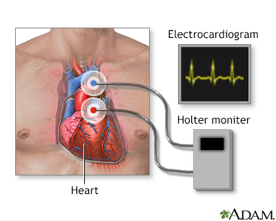
Holter heart monitor - illustration
During a heart Holter monitor study, the patient wears a monitor that records electrical activity of their heart (similarly to the recording of an electrocardiogram). This usually occurs for 24 hours, while at the same time the patient also records a diary of their activity. Health care providers then analyze the recording, tabulate a report of the heart's activity, and correlate irregular heart activity with the entries of the patient's diary.
Holter heart monitor
illustration
-
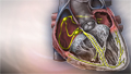
Heart - section through the middle - illustration
The interior of the heart is composed of valves, chambers, and associated vessels.
Heart - section through the middle
illustration
-
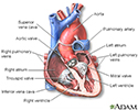
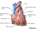
Heart - front view - illustration
The external structures of the heart include the ventricles, atria, arteries and veins. Arteries carry blood away from the heart while veins carry blood into the heart. The vessels colored blue indicate the transport of blood with relatively low content of oxygen and high content of carbon dioxide. The vessels colored red indicate the transport of blood with relatively high content of oxygen and low content of carbon dioxide.
Heart - front view
illustration
-
Normal heart rhythm - illustration
An electrocardiogram (ECG) test measures the electrical activity of the heart. A normal resting heart rate is 60 to 100 beats per minute.
Normal heart rhythm
illustration
-
Conduction system of the heart - illustration
The intrinsic conduction system sets the basic rhythm of the beating heart by generating impulses which stimulate the heart to contract.
Conduction system of the heart
illustration


-
Holter heart monitor - illustration
During a heart Holter monitor study, the patient wears a monitor that records electrical activity of their heart (similarly to the recording of an electrocardiogram). This usually occurs for 24 hours, while at the same time the patient also records a diary of their activity. Health care providers then analyze the recording, tabulate a report of the heart's activity, and correlate irregular heart activity with the entries of the patient's diary.
Holter heart monitor
illustration
-
Heart - section through the middle - illustration
The interior of the heart is composed of valves, chambers, and associated vessels.
Heart - section through the middle
illustration
-
Heart - front view - illustration
The external structures of the heart include the ventricles, atria, arteries and veins. Arteries carry blood away from the heart while veins carry blood into the heart. The vessels colored blue indicate the transport of blood with relatively low content of oxygen and high content of carbon dioxide. The vessels colored red indicate the transport of blood with relatively high content of oxygen and low content of carbon dioxide.
Heart - front view
illustration
-
Normal heart rhythm - illustration
An electrocardiogram (ECG) test measures the electrical activity of the heart. A normal resting heart rate is 60 to 100 beats per minute.
Normal heart rhythm
illustration
-
Conduction system of the heart - illustration
The intrinsic conduction system sets the basic rhythm of the beating heart by generating impulses which stimulate the heart to contract.
Conduction system of the heart
illustration
Review Date: 5/27/2024
Reviewed By: Michael A. Chen, MD, PhD, Associate Professor of Medicine, Division of Cardiology, Harborview Medical Center, University of Washington Medical School, Seattle, WA. Also reviewed by David C. Dugdale, MD, Medical Director, Brenda Conaway, Editorial Director, and the A.D.A.M. Editorial team.
 All rights reserved.