

Nuclear ventriculography
Cardiac blood pooling imaging; Heart scan - nuclear; Radionuclide ventriculography (RNV); Multiple gate acquisition scan (MUGA); Nuclear cardiology; Cardiomyopathy - nuclear ventriculographyNuclear ventriculography is a test that uses radioactive materials called tracers to show the heart chambers. The procedure is noninvasive. The instruments do not directly touch the heart.
Noninvasive
The term noninvasive can refer to diseases, procedures, or devices. Noninvasive diseases usually do not spread to or damage other organs and tissues...

How the Test is Performed
The test is done while you are resting.
The health care provider will inject a small amount of radioactive material called technetium into your vein. This substance attaches to red blood cells and passes through the heart.
The red blood cells inside the heart that carry the material form an image that a special camera can pick up. These scanners trace the substance as it moves through the heart area. The camera is timed with an electrocardiogram. A computer then processes the images to make it appear as if the heart is moving.
Electrocardiogram
An electrocardiogram (ECG) is a test that records the electrical activity of the heart.

How to Prepare for the Test
You may be told not to eat or drink for several hours before the test.
How the Test will Feel
You may feel a brief sting or pinch when the IV is inserted into your vein. Most often, a vein in the arm is used. You may have trouble staying still during the test.

Why the Test is Performed
The test will show how well the blood is pumping through different parts of the heart.
Normal Results
Normal results show that the heart squeezing function is normal. The test can check the overall squeezing strength of the heart (ejection fraction). A normal value is above 50% to 55%.
The test also can check the motion of different parts of the heart. If one part of the heart is moving poorly while the others move well, it may mean that there has been damage to that part of the heart.
What Abnormal Results Mean
Abnormal results may be due to:
- Blockages in the coronary arteries (coronary artery disease)
Coronary artery disease
Coronary heart disease is a narrowing of the blood vessels that supply blood and oxygen to the heart. Coronary heart disease (CHD) is also called co...
 ImageRead Article Now Book Mark Article
 - Heart valve disease
- Other cardiac disorders that weaken the heart muscle (reduced pumping function)
- Past heart attack (myocardial infarction)
Myocardial infarction
Most heart attacks are caused by a blood clot that blocks one of the coronary arteries. The coronary arteries bring blood and oxygen to the heart. ...
 ImageRead Article Now Book Mark Article
The test may also be performed for:
-
Dilated cardiomyopathy
Dilated cardiomyopathy
Cardiomyopathy is disease in which the heart muscle becomes weakened, stretched, or has another structural problem. Dilated cardiomyopathy is a condi...
 ImageRead Article Now Book Mark Article
 -
Heart failure
Heart failure
Heart failure is a condition in which the heart is no longer able to pump oxygen-rich blood to the rest of the body efficiently. This causes symptom...
 ImageRead Article Now Book Mark Article
 - Idiopathic cardiomyopathy
-
Peripartum cardiomyopathy
Peripartum cardiomyopathy
Peripartum cardiomyopathy is a rare disorder in which a pregnant woman's heart becomes weakened and enlarged. It develops during the last month of p...
ImageRead Article Now Book Mark Article - Ischemic cardiomyopathy
- Testing whether a medicine has affected heart function
Risks
Nuclear imaging tests carry a very low risk. Exposure to the radioisotope delivers a small amount of radiation. This amount is safe for people who do not have nuclear imaging tests often.
References
Bogaert J, Symons R. Ischaemic heart disease. In: Adam A, Dixon AK, Gillard JH, Schaefer-Prokop CM, eds. Grainger & Allison's Diagnostic Radiology: A Textbook of Medical Imaging. 7th ed. Philadelphia, PA: Elsevier; 2021:chap 15.
Dorbala S, Di Carli MF. Nuclear cardiology. In: Libby P, Bonow RO, Mann DL, Tomaselli GF, Bhatt DL, Solomon SD, eds. Braunwald's Heart Disease: A Textbook of Cardiovascular Medicine. 12th ed. Philadelphia, PA: Elsevier; 2022:chap 18.
Kramer CM, Dilsizian V, Hagspiel KD. Noninvasive cardiac imaging. In: Goldman L, Cooney KA, eds. Goldman-Cecil Medicine. 27th ed. Philadelphia, PA: Elsevier; 2024:chap 44.
Mettler FA, Guiberteau MJ. Cardiovascular system. In: Mettler FA, Guiberteau MJ, eds. Essentials of Nuclear Medicine and Molecular Imaging. 7th ed. Philadelphia, PA: Elsevier; 2019:chap 5.
-
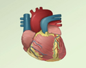
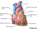
Heart - front view - illustration
The external structures of the heart include the ventricles, atria, arteries and veins. Arteries carry blood away from the heart while veins carry blood into the heart. The vessels colored blue indicate the transport of blood with relatively low content of oxygen and high content of carbon dioxide. The vessels colored red indicate the transport of blood with relatively high content of oxygen and low content of carbon dioxide.
Heart - front view
illustration
-
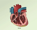
MUGA test - illustration
During the MUGA test, a radioactive isotope is injected into the vein. Radioactive isotopes attach to red blood cells and pass through the heart in the circulation. The isotopes can be traced through the heart using special cameras or scanners. The test is often given at rest, then repeated with exercise, or after administering certain medications. The test is performed to detect certain heart conditions.
MUGA test
illustration

-
Heart - front view - illustration
The external structures of the heart include the ventricles, atria, arteries and veins. Arteries carry blood away from the heart while veins carry blood into the heart. The vessels colored blue indicate the transport of blood with relatively low content of oxygen and high content of carbon dioxide. The vessels colored red indicate the transport of blood with relatively high content of oxygen and low content of carbon dioxide.
Heart - front view
illustration
-
MUGA test - illustration
During the MUGA test, a radioactive isotope is injected into the vein. Radioactive isotopes attach to red blood cells and pass through the heart in the circulation. The isotopes can be traced through the heart using special cameras or scanners. The test is often given at rest, then repeated with exercise, or after administering certain medications. The test is performed to detect certain heart conditions.
MUGA test
illustration
Review Date: 5/8/2024
Reviewed By: Thomas S. Metkus, MD, Assistant Professor of Medicine and Surgery, Johns Hopkins University School of Medicine, Baltimore, MD. Also reviewed by David C. Dugdale, MD, Medical Director, Brenda Conaway, Editorial Director, and the A.D.A.M. Editorial team.
 All rights reserved.