

Joint x-ray
X-ray - joint; Arthrography; ArthrogramThis test is an x-ray of a knee, shoulder, hip, wrist, ankle, or other joint.
How the Test is Performed
The test is done in a hospital radiology department or in the health care provider's office. The x-ray technologist will help you position the joint to be x-rayed on the table. Once in place, pictures are taken. The joint may be moved into other positions for more images.
How to Prepare for the Test
Tell your provider if you are pregnant. Remove all jewelry before the x-ray.
How the Test will Feel
The x-ray is painless. It may be uncomfortable to move the joint into different positions.
x-ray
X-rays are a type of electromagnetic radiation, just like visible light. An x-ray machine sends individual x-ray waves through the body. The images...

Why the Test is Performed
The x-ray is used to detect fractures, tumors, or degenerative conditions of the joint.
Fractures
If more pressure is put on a bone than it can stand, it will split or break. A break of any size is called a fracture. If the broken bone punctures...
What Abnormal Results Mean
The x-ray may show:
- Arthritis
Arthritis
Arthritis is inflammation or degeneration of one or more joints. A joint is the area where 2 bones meet. There are more than 100 different types of...
 ImageRead Article Now Book Mark Article
 - Fractures
- Bone tumors
Bone tumors
A bone tumor is an abnormal growth of cells within a bone. A bone tumor may be cancerous (malignant) or noncancerous (benign).
ImageRead Article Now Book Mark Article - Degenerative bone conditions
- Osteomyelitis (infection of the bone)
Osteomyelitis
Osteomyelitis is a bone infection. It is caused by bacteria or other germs.
ImageRead Article Now Book Mark Article
The test may also be performed to find out more about the following conditions:
- Acute gouty arthritis (gout)
Acute gouty arthritis (gout)
Gout is a type of arthritis. It occurs when uric acid builds up in the blood and causes inflammation in the joints. Acute gout is a painful conditio...
 ImageRead Article Now Book Mark Article
 - Adult-onset Still disease
Adult-onset Still disease
Adult Still disease (ASD) is a rare illness that causes high fevers, rash, and joint pain. It may lead to long-term (chronic) arthritis. Adult Still...
Read Article Now Book Mark Article - Caplan syndrome
Caplan syndrome
Rheumatoid pneumoconiosis (RP, also known as Caplan syndrome) is swelling (inflammation) and scarring of the lungs. It occurs in people with rheumat...
 ImageRead Article Now Book Mark Article
 - Chondromalacia patellae
Chondromalacia patellae
Anterior knee pain is pain that occurs at the front and center of the knee. It can be caused by many different problems, including:Chondromalacia of...
 ImageRead Article Now Book Mark Article
 - Chronic gouty arthritis
- Congenital dislocation of the hip
Congenital dislocation of the hip
Developmental dysplasia of the hip (DDH) is a dislocation of the hip joint that is present at birth. The condition is found in babies or young child...
 ImageRead Article Now Book Mark Article
 - Fungal arthritis
Fungal arthritis
Fungal arthritis is swelling and irritation (inflammation) of a joint by a fungal infection. It is also called mycotic arthritis.
 ImageRead Article Now Book Mark Article
 - Non-gonococcal (septic) bacterial arthritis
Non-gonococcal (septic) bacterial arthr...
Septic arthritis is inflammation of a joint due to a bacterial or fungal infection. Septic arthritis that is due to the bacteria that cause gonorrhe...
 ImageRead Article Now Book Mark Article
 - Osteoarthritis
Osteoarthritis
Osteoarthritis (OA) is the most common joint disorder. It is due to aging and wear and tear on a joint.
 ImageRead Article Now Book Mark Article
 - Pseudogout
Pseudogout
Calcium pyrophosphate dihydrate (CPPD) arthritis is a joint disease that can cause attacks of arthritis. Like gout, crystals form in the joints. Bu...
 ImageRead Article Now Book Mark Article
 - Psoriatic arthritis
Psoriatic arthritis
Psoriatic arthritis is a joint problem (arthritis) that often occurs with a skin condition called psoriasis.
 ImageRead Article Now Book Mark Article
 - Reiter syndrome
Reiter syndrome
Reactive arthritis is a type of arthritis that follows an infection. It may also cause inflammation of the eyes, skin and urinary and genital system...
 ImageRead Article Now Book Mark Article
 - Rheumatoid arthritis
Rheumatoid arthritis
Rheumatoid arthritis (RA) is a disease that leads to inflammation of the joints and surrounding tissues. It is a long-term disease. It can also aff...
 ImageRead Article Now Book Mark Article
 - Runner's knee
Runner's knee
Anterior knee pain is pain that occurs at the front and center of the knee. It can be caused by many different problems, including:Chondromalacia of...
ImageRead Article Now Book Mark Article - Tuberculous arthritis
Risks
There is low radiation exposure. X-ray machines are set to provide the smallest amount of radiation exposure needed to produce the image. Most experts feel that the risk is low compared with the benefits. Children and the fetuses of pregnant women are more sensitive to the risks of the x-ray. A protective shield may be worn over areas not being scanned.
References
Contreras F, Perez J, Jose J. Imaging overview. In: Miller MD, Thompson SR, eds. DeLee, Drez, & Miller's Orthopaedic Sports Medicine. 5th ed. Philadelphia, PA: Elsevier; 2020:chap 7.
Kapoor G, Toms AP. Current status of imaging of the musculoskeletal system. In: Adam A, Dixon AK, Gillard JH, Schaefer-Prokop CM, eds. Grainger & Allison's Diagnostic Radiology: A Textbook of Medical Imaging. 7th ed. Philadelphia, PA: Elsevier; 2021:chap 38.
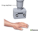
Hand X-ray - illustration
An x-ray is a photo taken with a machine which passes electromagnetic radiation through the body, capturing an image of the internal structures.
Hand X-ray
illustration
Review Date: 4/27/2023
Reviewed By: Linda J. Vorvick, MD, Clinical Professor, Department of Family Medicine, UW Medicine, School of Medicine, University of Washington, Seattle, WA. Also reviewed by David C. Dugdale, MD, Medical Director, Brenda Conaway, Editorial Director, and the A.D.A.M. Editorial team.
 All rights reserved.