Bone x-ray
X-ray - boneA bone x-ray is an imaging test to look at the bones.
x-ray
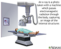
X-rays are a type of electromagnetic radiation, just like visible light. An x-ray machine sends individual x-ray waves through the body. The images...

How the Test is Performed
The test is done in a hospital radiology department or in your health care provider's office by an x-ray technician. For the test, you will position the bone to be x-rayed on the table. Pictures are then taken, and the bone is repositioned for different views.
How to Prepare for the Test
Tell your health care provider if you are pregnant. You must remove all jewelry for the x-ray.
How the Test will Feel
The x-rays are painless. Changing position for getting different views of the bone may be uncomfortable.
Why the Test is Performed
A bone x-ray is used to look for injuries or conditions affecting the bone.
What Abnormal Results Mean
Abnormal findings include:
- Fractures or broken bone
-
Bone tumors
Bone tumors
A bone tumor is an abnormal growth of cells within a bone. A bone tumor may be cancerous (malignant) or noncancerous (benign).
ImageRead Article Now Book Mark Article - Degenerative bone conditions
-
Osteomyelitis (bone infection)
Osteomyelitis
Osteomyelitis is a bone infection. It is mainly caused by bacteria or other germs.
ImageRead Article Now Book Mark Article
Additional conditions under which the test may be performed:
-
Cystic fibrosis
Cystic fibrosis
Cystic fibrosis is a disease that causes thick, sticky mucus to build up in the lungs, digestive tract, and other areas of the body. It is one of th...
 ImageRead Article Now Book Mark Article
 -
Multiple endocrine neoplasia (MEN) II
Multiple endocrine neoplasia
Multiple endocrine neoplasia, type II (MEN II) is a disorder passed down through families in which one or more of the endocrine glands are overactive...
 ImageRead Article Now Book Mark Article
 -
Multiple myeloma
Multiple myeloma
Multiple myeloma is a blood cancer that starts in the plasma cells in the bone marrow. Bone marrow is the soft, spongy tissue found inside most bone...
 ImageRead Article Now Book Mark Article
 -
Osgood-Schlatter disease
Osgood-Schlatter disease
Osgood-Schlatter disease is a painful swelling of the bump on the upper part of the shinbone, just below the knee. This bump is called the anterior ...
 ImageRead Article Now Book Mark Article
 -
Osteogenesis imperfecta
Osteogenesis imperfecta
Osteogenesis imperfecta is a condition causing extremely fragile bones.
Read Article Now Book Mark Article -
Osteomalacia
Osteomalacia
Osteomalacia is softening of the bones. It most often occurs because of a problem with vitamin D, which helps your body absorb calcium. Your body n...
 ImageRead Article Now Book Mark Article
 -
Paget's disease
Paget's disease
Paget disease is a disorder that involves abnormal bone destruction and regrowth. This results in deformity of the affected bones.
ImageRead Article Now Book Mark Article - Primary hyperparathyroidism
-
Rickets
Rickets
Rickets is a disorder caused by a lack of vitamin D, calcium, or phosphate. It leads to softening and weakening of the bones.
ImageRead Article Now Book Mark Article
Risks
There is low radiation exposure. X-ray machines are set to provide the smallest amount of radiation exposure needed to produce the image. Most experts feel that the risk is low compared with the benefits.
Children and the fetuses of pregnant women are more sensitive to the risks of the x-ray. A protective shield may be worn over areas not being scanned.
References
Contreras F, Perez J, Jose J. Imaging overview. In: Miller MD, Thompson SR. eds. DeLee, Drez, & Miller's Orthopaedic Sports Medicine. 5th ed. Philadelphia, PA: Elsevier; 2020:chap 7.
Kapoor G, Toms AP. Current status of imaging of the musculoskeletal system. In: Adam A, Dixon AK, Gillard JH, Schaefer-Prokop CM, eds. Grainger & Allison's Diagnostic Radiology: A Textbook of Medical Imaging. 7th ed. Philadelphia, PA: Elsevier; 2021:chap 38.
-
Skeleton - illustration
The skeleton consists of groups of bones which protect and move the body.
Skeleton
illustration
-
Skeletal spine - illustration
The spine is divided into several sections. The cervical vertebrae make up the neck. The thoracic vertebrae comprise the chest section and have ribs attached. The lumbar vertebrae are the remaining vertebrae below the last thoracic bone and the top of the sacrum. The sacral vertebrae are caged within the bones of the pelvis, and the coccyx represents the terminal vertebrae or vestigial tail.
Skeletal spine
illustration
-
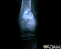
Osteogenic sarcoma - X-ray - illustration
This X-ray shows a malignant bone tumor (osteogenic sarcoma) of the knee. This type of tumor is usually seen in adolescents (around 15 years old). This tumor extends from the bone into the surrounding tissue.
Osteogenic sarcoma - X-ray
illustration
-
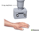
Hand X-ray - illustration
An x-ray is a photo taken with a machine which passes electromagnetic radiation through the body, capturing an image of the internal structures.
Hand X-ray
illustration
-
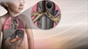
X-ray - illustration
X-rays are a form of ionizing radiation that can penetrate the body to form an image on film. Structures that are dense (such as bone) will appear white, air will be black, and other structures will be shades of gray depending on density. X-rays can provide information about obstructions, tumors, and other diseases, especially when coupled with the use of barium and air contrast within the bowel.
X-ray
illustration


-
Skeleton - illustration
The skeleton consists of groups of bones which protect and move the body.
Skeleton
illustration
-
Skeletal spine - illustration
The spine is divided into several sections. The cervical vertebrae make up the neck. The thoracic vertebrae comprise the chest section and have ribs attached. The lumbar vertebrae are the remaining vertebrae below the last thoracic bone and the top of the sacrum. The sacral vertebrae are caged within the bones of the pelvis, and the coccyx represents the terminal vertebrae or vestigial tail.
Skeletal spine
illustration
-
Osteogenic sarcoma - X-ray - illustration
This X-ray shows a malignant bone tumor (osteogenic sarcoma) of the knee. This type of tumor is usually seen in adolescents (around 15 years old). This tumor extends from the bone into the surrounding tissue.
Osteogenic sarcoma - X-ray
illustration
-
Hand X-ray - illustration
An x-ray is a photo taken with a machine which passes electromagnetic radiation through the body, capturing an image of the internal structures.
Hand X-ray
illustration
-
X-ray - illustration
X-rays are a form of ionizing radiation that can penetrate the body to form an image on film. Structures that are dense (such as bone) will appear white, air will be black, and other structures will be shades of gray depending on density. X-rays can provide information about obstructions, tumors, and other diseases, especially when coupled with the use of barium and air contrast within the bowel.
X-ray
illustration
Review Date: 4/27/2023
Reviewed By: Linda J. Vorvick, MD, Clinical Professor, Department of Family Medicine, UW Medicine, School of Medicine, University of Washington, Seattle, WA. Also reviewed by David C. Dugdale, MD, Medical Director, Brenda Conaway, Editorial Director, and the A.D.A.M. Editorial team.
 All rights reserved.