Eye and orbit ultrasound
An eye and orbit ultrasound is a test to look at the eye area. It also measures the size and structures of the eye.
Ultrasound
Ultrasound uses high-frequency sound waves to make images of organs and structures inside the body.

How the Test is Performed
The test is most often done in the ophthalmologist's office or the ophthalmology department of a hospital or clinic.
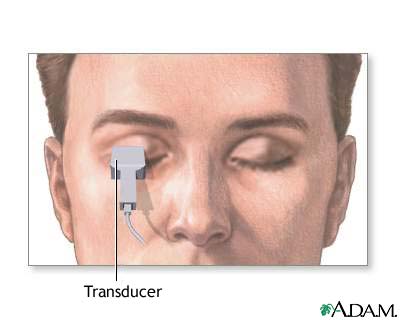
Your eye is numbed with anesthetic drops. The ultrasound wand (transducer) is placed against the front surface of the eye.
The ultrasound uses high-frequency sound waves that travel through the eye. Reflections (echoes) of the sound waves form a picture of the structure of the eye. The test takes about 15 minutes.
There are two types of scans: A-scan and B-scan.
For the A-scan:
- You will most often sit in a chair and place your chin on a chin rest. You will look straight ahead.
- A small probe is placed against the front of your eye.
- The test may also be done with you lying back. With this method, a fluid-filled cup is placed against your eye to do the test.
For the B-scan:
- You will be seated and you may be asked to look in many directions. The test is most often done with your eyes closed.
- A gel is placed on the skin of your eyelids. The B-scan probe is gently placed against your eyelids to do the test.
How to Prepare for the Test
No special preparation is needed for this test.
How the Test will Feel
Your eye is numbed, so you should not have any discomfort. You may be asked to look in different directions to improve the ultrasound image or so it can view different areas of your eye.
The gel used with the B-scan may run down your cheek, but you will not feel any discomfort or pain.
Why the Test is Performed
You may need this test if you have cataracts or other eye problems.
An A-scan ultrasound measures the eye to determine the right power of a lens implant before cataract surgery.
Cataract surgery
Cataract removal is surgery to remove a clouded lens (cataract) from the eye. Cataracts are removed to help you see better. The procedure almost al...

A B-scan is done to look at the inside part of the eye or the space around and behind the eye (orbit) that cannot be seen directly. This may occur when you have cataracts or other conditions that make it hard for the doctor to see into the back of your eye. The test may help diagnose retinal detachment, tumors, or other disorders.
Retinal detachment
Retinal detachment is a separation of the light-sensitive membrane (retina) in the back of the eye from its supporting layers.
Normal Results
For an A-scan, measurements of the eye are in the normal range.
For a B-scan, the structures of the eye and orbit appear normal.
What Abnormal Results Mean
A B-scan may show:
- Bleeding into the clear gel (vitreous) that fills the back of the eye (vitreous hemorrhage)
- Cancer of the retina (retinoblastoma), under the retina, or in other parts of the eye (such as melanoma)
Retinoblastoma
Retinoblastoma is a rare eye tumor that usually occurs in children. It is a malignant (cancerous) tumor of the part of the eye called the retina....
ImageRead Article Now Book Mark ArticleMelanoma
Melanoma of the eye is cancer that occurs in various parts of the eye.
 ImageRead Article Now Book Mark Article
 - Damaged tissue or injuries in the bony socket (orbit) that surrounds and protects the eye
- Foreign bodies
- Pulling away of the retina from the back of the eye (retinal detachment)
Retinal detachment
Retinal detachment is a separation of the light-sensitive membrane (retina) in the back of the eye from its supporting layers.
ImageRead Article Now Book Mark Article - Swelling (inflammation)
Risks
To avoid scratching the cornea, do not rub the numbed eye until the anesthetic wears off (about 15 minutes). There are no other risks.
Reviewed By
Franklin W. Lusby, MD, Ophthalmologist, Lusby Vision Institute, La Jolla, CA. Also reviewed by David C. Dugdale, MD, Medical Director, Brenda Conaway, Editorial Director, and the A.D.A.M. Editorial team.
Campion T, Miszkiel K, Davagnanam I. The orbit. In: Adam A, Dixon AK, Gillard JH, Schaefer-Prokop CM, eds. Grainger & Allison's Diagnostic Radiology. 7th ed. Philadelphia, PA: Elsevier; 2021:chap 60.
Fisher YL, Bacci T. Contact B-scan ultrasonography. In: Yanoff M, Duker JS, eds. Ophthalmology. 6th ed. Philadelphia, PA: Elsevier; 2023:chap 6.3.
Fisher YL, Silverman RH, Ledesma-Gill G, Engelbert M. Diagnostic ophthalmic ultrasound. In: Sadda SVR, Sarraf D, Freund KB, et al, eds. Ryan's Retina. 7th ed. Philadelphia, PA: Elsevier; 2023:chap 10.
 All rights reserved.