

Heart MRI
Magnetic resonance imaging - cardiac; Magnetic resonance imaging - heart; Nuclear magnetic resonance - cardiac; NMR - cardiac; MRI of the heart; Cardiomyopathy - MRI; Heart failure - MRI; Congenital heart disease - MRIHeart magnetic resonance imaging is an imaging method that uses powerful magnets and radio waves to create pictures of the heart. It does not use radiation (x-rays).
Magnetic resonance imaging
A magnetic resonance imaging (MRI) scan is an imaging test that uses powerful magnets and radio waves to create pictures of the body. It does not us...

Single magnetic resonance imaging (MRI) images are called slices. The images can be stored on a computer or printed on film. One exam produces dozens or sometimes hundreds of images.
The test may be done as part of a chest MRI.
Chest MRI
A chest MRI (magnetic resonance imaging) scan is an imaging test that uses powerful magnetic fields and radio waves to create pictures of the chest (...
How the Test is Performed
You may be asked to wear a hospital gown or clothing without metal fasteners (such as sweatpants and a t-shirt). Some types of metal can cause blurry images or be attracted to the powerful magnet.
You will lie on a narrow table, which slides into a large tunnel-like tube.
Some exams require a special dye (contrast). The dye is most often given before the test through a vein (IV) in your hand or forearm. The dye helps the radiologist see certain areas more clearly. This is different from the dye used for a CT scan.
During the MRI, the person who operates the machine will watch you from another room. The test most often lasts 30 to 60 minutes but may take longer.
How to Prepare for the Test
You may be asked not to eat or drink anything for 4 to 6 hours before the scan.
Tell your health care provider if you are afraid of close spaces (have claustrophobia). You may be given a medicine to help you feel sleepy and less anxious, or your provider may suggest an "open" MRI, in which the machine is not as close to the body.
Before the test, tell your provider if you have:
- Brain aneurysm clips
- Certain types of artificial heart valves
- Heart defibrillator or pacemaker
- Inner ear (cochlear) implants
- Kidney disease or dialysis (you may not be able to receive contrast)
- Recently placed artificial joints
- Certain types of vascular stents
Stents
A stent is a tiny tube placed into a hollow structure in your body. This structure can be an artery, a vein, or another structure, such as the tube ...
 ImageRead Article Now Book Mark Article
 - Worked with sheet metal in the past (you may need tests to check for metal pieces in your eyes)
Because the MRI contains strong magnets, metal objects are not allowed into the room with the MRI scanner:
- Pens, pocketknives, and eyeglasses may fly across the room.
- Items such as jewelry, watches, credit cards, and hearing aids can be damaged.
- Pins, hairpins, metal zippers, and similar metallic items can distort the images.
- Removable dental work should be taken out just before the scan.
How the Test will Feel
A heart MRI exam causes no pain. Some people may become anxious when inside the scanner. If you have a hard time lying still or are very anxious, you may be given medicine to relax. Too much movement can blur MRI images and cause errors.
The table may be hard or cold, but you can ask for a blanket or pillow. The machine produces loud thumping and humming noises when turned on. You may be given ear plugs to help reduce the noise.
An intercom in the scanner allows you to speak to the person operating the exam at any time. Some MRI scanners have televisions and special headphones to help pass the time.
There is no recovery time, unless sedation was necessary. (You will need someone to drive you home if sedation was given.) After an MRI scan, you can resume your normal diet, activity, and medicines, unless your provider tells you otherwise.
Why the Test is Performed
MRI provides detailed pictures of the heart and blood vessels from many views. Often, it is used when more information is needed after you have had an echocardiogram or heart CT scan. MRI is more accurate than CT scan or other tests for certain conditions, but less accurate for others.
Heart MRI may be used to evaluate or diagnose:
- Heart muscle damage after a heart attack
- Birth defects of the heart
- Heart tumors and growths
- Weakening or other problems with the heart muscle
- Symptoms of heart failure
- Valve disease or shunts
What Abnormal Results Mean
Abnormal results may be due to many things, including:
- Heart valve disorders
- Fluid in the sac-like covering around the heart (pericardial effusion)
Pericardial effusion
Pericarditis is a condition in which the sac-like covering around the heart (pericardium) becomes inflamed.
 ImageRead Article Now Book Mark Article
 - Tumor of the blood vessels or around the heart
-
Atrial myxoma or another growth or tumor in the heart
Atrial myxoma
An atrial myxoma is a noncancerous tumor in the upper left or right side of the heart. It most often grows on the wall that separates the two sides ...
 ImageRead Article Now Book Mark Article
 - Congenital heart disease (heart problem that you are born with)
- Damage or death to the heart muscle, seen after a heart attack
Heart attack
Most heart attacks are caused by a blood clot that blocks one of the coronary arteries. The coronary arteries bring blood and oxygen to the heart. ...
 ImageRead Article Now Book Mark Article
 - Inflammation of the heart muscle (myocarditis)
- Infiltration of the heart muscle by unusual substances
- Weakening of the heart muscle, which can be caused by sarcoidosis or amyloidosis
Risks
There is no radiation involved in MRI. The magnetic fields and radio waves used during the scan have not been shown to cause any significant side effects.
Allergic reactions to the dye used during the exam are rare. The most common type of contrast (dye) used is gadolinium. It is very safe. The person operating the machine will monitor your heart rate and breathing as needed. Rare complications can occur in people with severe kidney problems.
People have been harmed in MRI machines when they did not remove metal objects from their clothes or when metal objects were left in the room by others.
MRI is most often not recommended for traumatic injuries. Traction and life-support equipment cannot safely enter the scanner area.
Traction
Traction means pulling on part of the body. Most often, traction uses devices such as weights and pulleys to put tension on a displaced bone or joint...
Read Article Now Book Mark ArticleMRIs can be costly, take a long time to perform, and are sensitive to movement.
References
Kramer CM, Dilsizian V, Hagspiel KD. Noninvasive cardiac imaging. In: Goldman L, Cooney KA, eds. Goldman-Cecil Medicine. 27th ed. Philadelphia, PA: Elsevier; 2024:chap 44.
Kwong RY. Cardiovascular magnetic resonance imaging. In: Libby P, Bonow RO, Mann DL, Tomaselli GF, Bhatt DL, Solomon SD, eds. Braunwald's Heart Disease: A Textbook of Cardiovascular Medicine. 12th ed. Philadelphia, PA: Elsevier; 2022:chap 19.
-
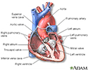
Heart - section through the middle - illustration
The interior of the heart is composed of valves, chambers, and associated vessels.
Heart - section through the middle
illustration
-
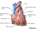
Heart - front view - illustration
The external structures of the heart include the ventricles, atria, arteries and veins. Arteries carry blood away from the heart while veins carry blood into the heart. The vessels colored blue indicate the transport of blood with relatively low content of oxygen and high content of carbon dioxide. The vessels colored red indicate the transport of blood with relatively high content of oxygen and low content of carbon dioxide.
Heart - front view
illustration
-
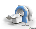
MRI scans - illustration
MRI stands for magnetic resonance imaging. It allows imaging of the interior of the body without using x-rays or other types of ionizing radiation. An MRI scan is capable of showing fine detail of different tissues.
MRI scans
illustration
-
Heart - section through the middle - illustration
The interior of the heart is composed of valves, chambers, and associated vessels.
Heart - section through the middle
illustration
-
Heart - front view - illustration
The external structures of the heart include the ventricles, atria, arteries and veins. Arteries carry blood away from the heart while veins carry blood into the heart. The vessels colored blue indicate the transport of blood with relatively low content of oxygen and high content of carbon dioxide. The vessels colored red indicate the transport of blood with relatively high content of oxygen and low content of carbon dioxide.
Heart - front view
illustration
-
MRI scans - illustration
MRI stands for magnetic resonance imaging. It allows imaging of the interior of the body without using x-rays or other types of ionizing radiation. An MRI scan is capable of showing fine detail of different tissues.
MRI scans
illustration
-
Heart failure - InDepth
(In-Depth)
Review Date: 2/27/2024
Reviewed By: Thomas S. Metkus, MD, Assistant Professor of Medicine and Surgery, Johns Hopkins University School of Medicine, Baltimore, MD. Also reviewed by David C. Dugdale, MD, Medical Director, Brenda Conaway, Editorial Director, and the A.D.A.M. Editorial team.
 All rights reserved.