Endocervical culture
Vaginal culture; Female genital tract culture; Culture - cervixEndocervical culture is a laboratory test that helps identify infection in the female genital tract.
How the Test is Performed
During a vaginal examination, the health care provider uses a swab to take samples of mucus and cells from the endocervix. This is the area around the opening of the uterus. The samples are sent to a lab. There, they are placed in a special dish (culture). They are then watched to see if bacteria, virus, or fungus grow. Further tests may be done to identify the specific organism and determine the best treatment.
How to Prepare for the Test
In the 2 days before the procedure:
- Do not use creams or other medicines in the vagina.
- Do not douche. (You should never douche. Douching can cause infection of the vagina or uterus.)
- Avoid intercourse.
- Empty your bladder and bowel.
- At your provider's office, follow instructions for preparing for the vaginal exam.
How the Test will Feel
You will feel some pressure from the speculum. This is an instrument inserted into the vagina to hold the area open so that the provider can view the cervix and collect the samples. There may be a slight cramping when the swab touches the cervix.
Cervix
The cervix is the lower end of the womb (uterus). It is at the top of the vagina. It is about 2. 5 to 3. 5 centimeters (1 to 1. 3 inches) long. Th...

Why the Test is Performed
The test may be done to determine the cause of vaginitis, pelvic pain, an unusual vaginal discharge, or other signs of infection.
Vaginitis
Vaginitis is a swelling or infection of the vulva and vagina. It may also be called vulvovaginitis. Vaginitis is a common problem that can affect wo...
Read Article Now Book Mark ArticleVaginal discharge
Vaginal discharge refers to secretions from the vagina. The discharge may be:Thick, pasty, or thinClear, cloudy, bloody, white, yellow, or greenOdor...

Normal Results
Organisms that are usually present in the vagina are there in the expected amounts.
What Abnormal Results Mean
Abnormal results indicate the presence of an infection in the genital tract or urinary tract in women, such as:
-
Genital herpes
Genital herpes
Genital herpes is a sexually transmitted infection. It is caused by the herpes simplex virus (HSV). This article focuses on HSV type 2 infection....
Read Article Now Book Mark Article - Chronic swelling and irritation of the urethra (urethritis)
Urethritis
Urethritis is inflammation (swelling and irritation) of the urethra. The urethra is the tube that carries urine from the body.
 ImageRead Article Now Book Mark Article
 - Sexually transmitted infections, such as gonorrhea or chlamydia
Gonorrhea
Gonorrhea is a common sexually transmitted infection (STI).
Read Article Now Book Mark ArticleChlamydia
Chlamydia is an infection caused by the bacteria Chlamydia trachomatis. It is most often spread through sexual contact.
 ImageRead Article Now Book Mark Article
 -
Pelvic inflammatory disease (PID)
Pelvic inflammatory disease
Pelvic inflammatory disease (PID) is an infection of a woman's womb (uterus), ovaries, or fallopian tubes.
 ImageRead Article Now Book Mark Article
Risks
There may be slight bleeding or spotting after the test. This is normal.
References
Eckert LO, Lentz GM. Genital tract infections: vulva, vagina, cervix, toxic shock syndrome, endometritis, and salpingitis. In: Gershenson DM, Lentz GM, Valea FA, Lobo RA, eds. Comprehensive Gynecology. 8th ed. Philadelphia, PA: Elsevier; 2022:chap 23.
Swygard H, Cohen MS. Approach to the patient with a sexually transmitted infection. In: Goldman L, Cooney KA, eds. Goldman-Cecil Medicine. 27th ed. Philadelphia, PA: Elsevier; 2024:chap 264.
-
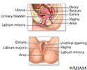
Female reproductive anatomy - illustration
Internal structures of the female reproductive anatomy include the uterus, ovaries, and cervix. External structures include the labium minora and majora, the vagina and the clitoris.
Female reproductive anatomy
illustration
-
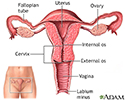
Uterus - illustration
The uterus is a hollow muscular organ located in the female pelvis between the bladder and rectum. The ovaries produce the eggs that travel through the fallopian tubes. Once the egg has left the ovary it can be fertilized and implant itself in the lining of the uterus. The main function of the uterus is to nourish the developing fetus prior to birth.
Uterus
illustration
-
Female reproductive anatomy - illustration
Internal structures of the female reproductive anatomy include the uterus, ovaries, and cervix. External structures include the labium minora and majora, the vagina and the clitoris.
Female reproductive anatomy
illustration
-
Uterus - illustration
The uterus is a hollow muscular organ located in the female pelvis between the bladder and rectum. The ovaries produce the eggs that travel through the fallopian tubes. Once the egg has left the ovary it can be fertilized and implant itself in the lining of the uterus. The main function of the uterus is to nourish the developing fetus prior to birth.
Uterus
illustration
Review Date: 8/23/2023
Reviewed By: LaQuita Martinez, MD, Department of Obstetrics and Gynecology, Emory Johns Creek Hospital, Alpharetta, GA. Also reviewed by David C. Dugdale, MD, Medical Director, Brenda Conaway, Editorial Director, and the A.D.A.M. Editorial team.


