Joint fluid Gram stain
Gram stain of joint fluidJoint fluid Gram stain is a lab test to identify bacteria in a sample of joint fluid using a special series of stains (colors). The Gram stain method is one of the most commonly used ways to quickly diagnose bacterial infections.
Gram stain method
A Gram stain is a test used to identify bacteria. It is one of the most common ways to quickly diagnose bacterial infection in the body.

How the Test is Performed
A sample of joint fluid is needed. This may be done in your health care provider's office using a needle, or during an operating room procedure. Removing the sample is called joint fluid aspiration.
Joint fluid aspiration
Synovial fluid analysis is a group of tests that examine joint (synovial) fluid. The tests help diagnose and treat joint-related problems.

The fluid sample is sent to a lab where a drop of fluid is spread in a very thin layer onto a microscope slide. This is called a smear. Several different colored stains are applied to the sample. The lab team member will look at the stained smear under a microscope to see if bacteria are present. The color, size, and shape of the cells help identify the bacteria.
How to Prepare for the Test
Your provider will tell you how to prepare for the procedure. No special preparation is needed. But, tell your provider if you're taking a blood thinner, such as aspirin, warfarin (Coumadin) or clopidogrel (Plavix). These medicines can affect test results or your ability to take the test.
How the Test will Feel
Sometimes, your provider will first inject numbing medicine into the skin with a small needle, which may sting. A larger needle is then used to draw out the synovial fluid.
This test may also cause some discomfort if the tip of the needle touches bone. The procedure usually lasts less than 1 to 2 minutes.
Why the Test is Performed
The test is performed when there is unexplained swelling, joint pain, and inflammation of a joint, or to check for suspected joint infection.

Normal Results
A normal result means no bacteria are present on the Gram stain. Other tests may be done to help diagnose the problem.
What Abnormal Results Mean
Abnormal results mean bacteria were seen on the Gram stain. This may be a sign of a joint infection, for example, gonococcal arthritis due to bacteria called Neisseria gonorrhoeae or arthritis due to bacteria called Staphylococcus aureus.
Gonococcal arthritis
Gonococcal arthritis is inflammation of a joint due to a gonorrhea infection. Gonococcal arthritis is a type of septic arthritis. This is inflammati...

Risks
Risks of this test include:
- Infection of the joint -- unusual, but more common with repeated aspirations
- Bleeding into the joint space
References
Karcher DS, McPherson RA. Cerebrospinal, synovial, serous body fluids, and alternative specimens. In: McPherson RA, Pincus MR, eds. Henry's Clinical Diagnosis and Management by Laboratory Methods. 24th ed. Philadelphia, PA: Elsevier; 2022:chap 30.
O'Neil L, Tanner S, El-Gabalawy HS. Synovial fluid analyses, synovial biopsy, and synovial pathology. In: Firestein GS, McInnes IB, Koretzky GA, Mikuls TR, Neogi T, O'Dell JR, eds. Firestein & Kelley's Textbook of Rheumatology. 12th ed. Philadelphia, PA: Elsevier; 2025:chap 53.
-
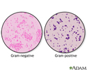
Gram stain - illustration
A Gram stain is a test used to help identify bacteria. The tested sample can be taken from body fluids that do not normally contain bacteria, such as blood, urine, or cerebrospinal fluid. A sample can also be taken from the site of a suspected infection, such as the throat, lungs, genitals, or skin. Bacteria are classified as either Gram-positive or Gram-negative, based on how they color in reaction with the Gram stain. The Gram stain is colored purple. When combined with the bacteria in a sample, the stain will either stay purple inside the bacteria (Gram-positive), or it will turn pink (Gram-negative). Examples of Gram-positive bacteria include Streptococcus and Staphylococcus aureus, as well as bacteria that cause anthrax, diphtheria, and toxic shock syndrome. Examples of Gram-negative bacteria include Escherichia coli (E coli), Salmonella, Hemophilus influenzae, as well as many bacteria that cause urinary tract infections, pneumonia, or peritonitis. Gram stain can be done within a few hours. The results help providers choose the first antibiotics to use. Cultures of bacteria help identify specific bacteria, but take days to complete.
Gram stain
illustration
-
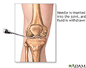
Joint aspiration - illustration
Synovial fluid analysis is a series of tests performed on synovial (joint) fluid to help diagnose and treat joint-related abnormalities. To obtain a synovial fluid sample, a needle is inserted into the knee between the joint space. When the needle is in place the synovial fluid is then withdrawn. The sample is sent to the lab for analysis.
Joint aspiration
illustration
-
Gram stain - illustration
A Gram stain is a test used to help identify bacteria. The tested sample can be taken from body fluids that do not normally contain bacteria, such as blood, urine, or cerebrospinal fluid. A sample can also be taken from the site of a suspected infection, such as the throat, lungs, genitals, or skin. Bacteria are classified as either Gram-positive or Gram-negative, based on how they color in reaction with the Gram stain. The Gram stain is colored purple. When combined with the bacteria in a sample, the stain will either stay purple inside the bacteria (Gram-positive), or it will turn pink (Gram-negative). Examples of Gram-positive bacteria include Streptococcus and Staphylococcus aureus, as well as bacteria that cause anthrax, diphtheria, and toxic shock syndrome. Examples of Gram-negative bacteria include Escherichia coli (E coli), Salmonella, Hemophilus influenzae, as well as many bacteria that cause urinary tract infections, pneumonia, or peritonitis. Gram stain can be done within a few hours. The results help providers choose the first antibiotics to use. Cultures of bacteria help identify specific bacteria, but take days to complete.
Gram stain
illustration
-
Joint aspiration - illustration
Synovial fluid analysis is a series of tests performed on synovial (joint) fluid to help diagnose and treat joint-related abnormalities. To obtain a synovial fluid sample, a needle is inserted into the knee between the joint space. When the needle is in place the synovial fluid is then withdrawn. The sample is sent to the lab for analysis.
Joint aspiration
illustration
-
Pneumonia - InDepth
(In-Depth)
-
Immunizations - InDepth
(In-Depth)
Review Date: 10/11/2024
Reviewed By: Frank D. Brodkey, MD, FCCM, Associate Professor, Section of Pulmonary and Critical Care Medicine, University of Wisconsin School of Medicine and Public Health, Madison, WI. Also reviewed by David C. Dugdale, MD, Medical Director, Brenda Conaway, Editorial Director, and the A.D.A.M. Editorial team.
 All rights reserved.