
Standard eye exam
Standard ophthalmic exam; Routine eye examination; Eye exam - standard; Annual eye examA standard eye exam is a series of tests done to check your vision and the health of your eyes.
How the Test is Performed
First, you will be asked if you are having any eye or vision problems. You will be asked to describe these problems, how long you have had them, and any factors that have made them better or worse.
Your history of glasses or contact lenses will also be reviewed. The eye doctor will then ask about your overall health, including any medicines you take and your family's medical history.
Next, the doctor will check your vision (visual acuity) using a Snellen chart.
Visual acuity
The visual acuity test is used to determine the smallest letters you can read on a standardized chart (Snellen chart) or a card held 20 feet (6 meter...

- You will be asked to read random letters that become smaller line by line as your eyes move down the chart. Some Snellen charts are actually video monitors showing letters or images.
- To see if you need glasses, the doctor will place several lenses in front of your eye, one at a time, and ask you when the letters on the Snellen chart become easier to see. This is called a refraction.
Refraction
A refraction is an eye exam that measures a person's prescription for eyeglasses or contact lenses.
 ImageRead Article Now Book Mark Article
Other parts of the exam include tests to:
- See if you have proper three-dimensional (3D) vision (stereopsis).
- Check your side (peripheral) vision.
- Check the eye muscles by asking you to look in different directions at a penlight or other small object.
- Examine the pupils with a penlight to see if they respond (constrict) properly to light.
- Often, you'll be given eye drops to open up (dilate) your pupils. This allows the doctor to use a device called an ophthalmoscope to view the structures at the back of the eye. This area is called the fundus. It includes the retina and nearby blood vessels and optic nerve.
Retina
The retina is the light-sensitive layer of tissue at the back of the eyeball. Images that come through the eye's lens are focused on the retina. Th...
ImageRead Article Now Book Mark Article
Another magnifying device, called a slit lamp, is used to:
Slit lamp
The slit-lamp examination looks at structures that are at the front of the eye.
- See the front parts of the eye (eyelids, cornea, conjunctiva, sclera, and iris)
- Check for increased pressure in the eye (glaucoma) using a method called tonometry
Glaucoma
Glaucoma is a group of eye conditions that can damage the optic nerve. This nerve sends the images you see to your brain. Most often, optic nerve da...
 ImageRead Article Now Book Mark Article
Tonometry
Tonometry is a test to measure the pressure inside your eyes. The test is used to screen for glaucoma. It is also used to measure how well glaucoma...
ImageRead Article Now Book Mark Article
Color blindness is tested using cards with colored dots that form numbers.
Color blindness
Color blindness is the inability to see some colors in the usual way.
Colored dots that form numbers
A color vision test checks your ability to distinguish between different colors.

How to Prepare for the Test
Make an appointment with an eye doctor (some take walk-in patients). Avoid eye strain on the day of the test. If you wear glasses or contacts, bring them with you. You may need someone to drive you home if the doctor uses eye drops to dilate your pupils.
How the Test will Feel
The tests cause no pain or discomfort.
Why the Test is Performed
All children should have vision screening in a pediatrician's or family practitioner's office around the time when they learn the alphabet, and then every 1 to 2 years afterward. Screening should begin sooner if any eye problems are suspected.
According to the American Academy of Ophthalmology, the following schedule should be used:
Between ages 20 and 39:
- A complete eye exam should be done every 5 to 10 years
- Adults who wear contact lenses need yearly eye exams
- Certain eye symptoms or disorders may require more frequent exams
Adults over age 40 who have no risk factors or ongoing eye conditions should be screened:
- Every 2 to 4 years for adults ages 40 to 54
- Every 1 to 3 years for adults ages 55 to 64
- Every 1 to 2 years for adults age 65 and older
Depending on your risk factors for eye diseases and your current symptoms or illnesses, your eye doctor may recommend that you have exams more often.
Some of the eye and medical problems that can be found by a routine eye test include:
- Cataracts (clouding of the lens of the eye)
- Diabetes
- Glaucoma
-
High blood pressure
High blood pressure
Blood pressure is a measurement of the force exerted against the walls of your arteries as your heart pumps blood to your body. Hypertension is the ...
 ImageRead Article Now Book Mark Article
 -
Age-related macular degeneration (ARMD), (loss of sharp, central vision)
Age-related macular degeneration
Macular degeneration is an eye disorder that slowly destroys sharp, central vision. This makes it difficult to see fine details and read. The diseas...
 ImageRead Article Now Book Mark Article
Normal Results
Results of a routine eye exam are normal when the eye doctor finds you have:
- 20/20 (6/6) (normal) vision
- Ability to identify different colors
- Full visual field
- Proper eye muscle coordination
- Normal eye pressure
- Normal eye structures (cornea, iris, lens)
What Abnormal Results Mean
Abnormal results may be due to any of the following:
- ARMD
-
Astigmatism (abnormally curved cornea)
Astigmatism
Astigmatism is a type of refractive error of the eye. Refractive errors cause blurred vision. They are the most common reason why a person goes to ...
 ImageRead Article Now Book Mark Article
 -
Blocked tear duct
Blocked tear duct
A blocked tear duct is a partial or complete blockage in the pathway that carries tears from the surface of the eye into the nose.
 ImageRead Article Now Book Mark Article
 - Cataracts
- Color blindness
-
Corneal dystrophy
Corneal dystrophy
Fuchs (pronounced "fooks") dystrophy is an eye disease in which cells lining the inner surface of the cornea slowly start to die off. The disease mo...
 ImageRead Article Now Book Mark Article
 -
Corneal ulcers, infections, or injury
Corneal ulcers, infections
The cornea is the clear tissue at the front of the eye. A corneal ulcer is an open sore in the outer layer of the cornea. It is often caused by inf...
ImageRead Article Now Book Mark ArticleInjury
Corneal injury is a wound to the part of the eye known as the cornea. The cornea is the crystal clear (transparent) tissue that covers the front of ...
ImageRead Article Now Book Mark Article - Damaged nerves or blood vessels in the eye
-
Diabetic retinopathy (Diabetes-related damage to the back of the eye)
Diabetic retinopathy
Diabetes can harm the eyes. It can damage the small blood vessels in the retina, the back part of your eye. This condition is called diabetic retin...
 ImageRead Article Now Book Mark Article
 -
Hyperopia (farsightedness)
Hyperopia
Farsightedness is having a harder time seeing objects that are close than things that are far away. The term is often used to describe the need for r...
 ImageRead Article Now Book Mark Article
 - Glaucoma
- Injury of the eye
-
Amblyopia (lazy eye)
Lazy eye
Amblyopia is the loss of the ability to see clearly through one eye. It is also called "lazy eye. " It is the most common cause of vision problems i...
ImageRead Article Now Book Mark Article -
Myopia (nearsightedness)
Myopia
Nearsightedness is when light entering the eye is focused incorrectly. This makes distant objects appear blurred. Nearsightedness is a type of refr...
ImageRead Article Now Book Mark Article -
Presbyopia (inability to focus on near objects that develops with age)
Presbyopia
Presbyopia is a condition in which the lens of the eye loses its ability to focus. This makes it hard to see objects up close.
 ImageRead Article Now Book Mark Article
 -
Strabismus (crossed eyes)
Strabismus
Strabismus is a disorder in which both eyes do not line up in the same direction. Therefore, they do not look at the same object at the same time. ...
 ImageRead Article Now Book Mark Article
 - Retinal tear or detachment
Detachment
Retinal detachment is a separation of the light-sensitive membrane (retina) in the back of the eye from its supporting layers.
ImageRead Article Now Book Mark Article
This list may not include all possible causes of abnormal results.
Risks
If you receive drops to dilate your eyes for the ophthalmoscopy, your vision will be blurred.
- Wear sunglasses to protect your eyes from sunlight, which can damage your eyes more when they are dilated.
- Have someone drive you home.
- The drops usually wear off in several hours.
In rare cases, the dilating eyedrops cause:
- An attack of narrow-angle glaucoma
- Dizziness
- Dryness of the mouth
- Flushing
- Nausea and vomiting
References
Ball JW, Dains JE, Flynn JA, Solomon BS, Stewart RW. Eyes. In: Ball JW, Dains JE, Flynn JA, Solomon BS, Stewart RW, eds. Seidel's Guide to Physical Examination. 10th ed. St Louis, MO: Elsevier; 2023:chap 12.
Chuck RS, Dunn SP, Flaxel CJ; American Academy of Ophthalmology Preferred Practice Pattern Committee, et al. Comprehensive adult medical eye evaluation preferred practice pattern. Ophthalmology. 2021;128(1):1-29. www.aaojournal.org/article/S0161-6420(20)31026-5/fulltext. Published November 12, 2020. Accessed April 5, 2023.
Olitsky SE, Marsh JD. Examination of the eye. In: Kliegman RM, St. Geme JW, Blum NJ, Shah SS, Tasker RC, Wilson KM, eds. Nelson Textbook of Pediatrics. 21st ed. Philadelphia, PA: Elsevier; 2020:chap 637.
Prokopich CL, Hrynchak P, Flanagan JG, Hynes AF, Chisholm C. Ocular health assessment. In: Elliott DB, ed. Clinical Procedures in Primary Eye Care. 5th ed. Philadelphia, PA: Elsevier; 2021:chap 7.
-
Visual acuity test - illustration
Visual acuity tests may be performed in many different ways. It is a quick way to detect vision problems and is frequently used in schools or for mass screening. Driver license bureaus often use a small device that can test the eyes both together and individually.
Visual acuity test
illustration
-
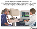
Visual field test - illustration
Central and peripheral vision is tested by using visual field tests. Changes may indicate eye diseases, such as glaucoma or retinitis.
Visual field test
illustration

-
Visual acuity test - illustration
Visual acuity tests may be performed in many different ways. It is a quick way to detect vision problems and is frequently used in schools or for mass screening. Driver license bureaus often use a small device that can test the eyes both together and individually.
Visual acuity test
illustration
-
Visual field test - illustration
Central and peripheral vision is tested by using visual field tests. Changes may indicate eye diseases, such as glaucoma or retinitis.
Visual field test
illustration
Review Date: 2/12/2023
Reviewed By: Franklin W. Lusby, MD, Ophthalmologist, Lusby Vision Institute, La Jolla, CA. Also reviewed by David C. Dugdale, MD, Medical Director, Brenda Conaway, Editorial Director, and the A.D.A.M. Editorial team.
 All rights reserved.