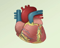
Cardiac catheterization
Catheterization - cardiac; Heart catheterization; Angina - cardiac catheterization; CAD - cardiac catheterization; Coronary artery disease - cardiac catheterization; Heart valve - cardiac catheterization; Heart failure - cardiac catheterizationCardiac catheterization involves passing a thin flexible tube (catheter) into the right or left side of the heart. The catheter is most often inserted from the groin or the arm.
How the Test is Performed
You will get medicine before the test to help you relax.
A member of the team doing this procedure will clean a site on your arm, neck, or groin and insert a small catheter into one of your veins. This is called an intravenous (IV) line.
A larger thin plastic tube called a sheath is placed into a vein or artery in your leg or arm. Then longer plastic tubes called catheters are carefully moved up into the heart using live x-rays (fluoroscopy) as a guide. Then the doctor (usually a cardiologist or heart specialist) doing the cardiac catheterization can:
x-rays
X-rays are a type of electromagnetic radiation, just like visible light. An x-ray machine sends individual x-ray waves through the body. The images...

- Collect blood samples from the heart
- Measure pressure and blood flow in the heart's chambers and in the large arteries around the heart
- Measure the oxygen levels in different parts of your heart
- Examine the arteries of the heart
- Perform a biopsy on the heart muscle
Biopsy on the heart muscle
Myocardial biopsy is the removal of a small piece of heart muscle for examination.
 ImageRead Article Now Book Mark Article
For some procedures, you may be injected with a dye that helps your doctor to visualize the structures and vessels within the heart.
If you have a blockage of one of the heart arteries, you may have angioplasty and a stent placed during the procedure.
The test may last 30 to 60 minutes. If you also need special procedures, the test may take longer. If the catheter is placed in your groin, you will often be asked to lie flat on your back for a few to several hours after the test to avoid bleeding.
You will be told how to take care of yourself when you go home after the procedure is done.
When you go home
Cardiac catheterization involves passing a thin flexible tube (catheter) into the right or left side of the heart. The catheter is most often insert...
Read Article Now Book Mark ArticleHow to Prepare for the Test
You should not eat or drink for 6 to 8 hours before the test. The test takes place in a hospital or specialized procedure unit and you will be asked to wear a hospital gown. Sometimes, you will need to spend the night before the test in the hospital. Otherwise, you will come to the hospital the morning of the procedure.
Your health care provider will explain the procedure and its risks. A witnessed, signed consent form for the procedure is required.
Tell your provider if you:
- Are allergic to seafood or any medicines
- Have had a bad reaction to contrast dye or iodine in the past
- Take any medicines, including Viagra or other medicines for erectile dysfunction
Erectile dysfunction
An erection problem occurs when a man cannot get or keep an erection that is firm enough for intercourse. You may not be able to get an erection at ...
 ImageRead Article Now Book Mark Article
 - Might be pregnant
How the Test will Feel
The test is done by cardiologists and a trained health care team.
You will be awake and able to follow instructions during the test.
You may feel some discomfort or pressure where the catheter is placed. You may have some discomfort from lying still during the test or from lying flat on your back after the procedure.
Why the Test is Performed
This procedure is most often done to get information about the heart or its blood vessels. It may also be done to treat some types of heart conditions, or to find out if you need heart surgery.
Your doctor may perform cardiac catheterization to diagnose or evaluate:
- Causes of congestive heart failure or cardiomyopathy
Congestive heart failure
Heart failure is a condition in which the heart is no longer able to pump oxygen-rich blood to the rest of the body efficiently. This causes symptom...
 ImageRead Article Now Book Mark Article
Cardiomyopathy
Cardiomyopathy is disease in which the heart muscle becomes weakened, stretched, or has another structural problem. It often contributes to the hear...
 ImageRead Article Now Book Mark Article
 - Coronary artery disease
Coronary artery disease
Coronary heart disease is a narrowing of the blood vessels that supply blood and oxygen to the heart. Coronary heart disease (CHD) is also called co...
 ImageRead Article Now Book Mark Article
 - Heart defects that are present at birth (congenital)
Heart defects
Congenital heart disease (CHD) is a problem with the heart's structure and function that is present at birth.
 ImageRead Article Now Book Mark Article
 - High blood pressure in the lungs (pulmonary hypertension)
Pulmonary hypertension
Pulmonary hypertension is high blood pressure in the pulmonary arteries of the lungs. It makes the right side of the heart work harder than normal....
 ImageRead Article Now Book Mark Article
 - Problems with the heart valves
The following procedures may also be done using cardiac catheterization:
- Repair certain types of heart defects
- Open a narrowed (stenotic) heart valve (valvuloplasty)
- Open blocked arteries or grafts in the heart (angioplasty with or without stenting)
Angioplasty
Angioplasty is a procedure to open narrowed or blocked blood vessels that supply blood to the heart. These blood vessels are called the coronary art...
 ImageRead Article Now Book Mark Article
Risks
Cardiac catheterization carries a slightly higher risk than other heart tests. However, it is very safe when done by an experienced team.
The risks include:
- Cardiac tamponade
Cardiac tamponade
Cardiac tamponade is pressure on the heart that occurs when blood or fluid builds up in the space between the heart muscle and the outer covering sac...
 ImageRead Article Now Book Mark Article
 - Heart attack
Heart attack
Most heart attacks are caused by a blood clot that blocks one of the coronary arteries. The coronary arteries bring blood and oxygen to the heart. ...
 ImageRead Article Now Book Mark Article
 - Injury to a coronary artery
- Irregular heartbeat
- Low blood pressure
Low blood pressure
Low blood pressure occurs when blood pressure is below normal. This means the heart, brain, and other parts of the body may not get enough blood. I...
 ImageRead Article Now Book Mark Article
 - Reaction to the contrast dye
- Stroke
Stroke
A stroke occurs when blood flow to a part of the brain stops. A stroke is sometimes called a "brain attack. " If blood flow is cut off for longer th...
 ImageRead Article Now Book Mark Article
Possible complications of any type of catheterization include the following:
- Bleeding, infection, and pain at the IV or sheath insertion site
- Damage to the blood vessels
- Blood clots
Blood clots
Blood clots are clumps that occur when blood hardens from a liquid to a solid. A blood clot that forms inside one of your veins or arteries is calle...
 ImageRead Article Now Book Mark Article
 - Kidney damage due to the contrast dye (more common in people with diabetes or kidney problems)
References
Dangas GD, Mehran R. Coronary angiography and intravascular imaging. In: Libby P, Bonow RO, Mann DL, Tomaselli GF, Bhatt DL, Solomon SD, eds. Braunwald's Heart Disease: A Textbook of Cardiovascular Medicine. 12th ed. Philadelphia, PA: Elsevier; 2022:chap 21.
Kumbhani DJ, Bhatt DL. Percutaneous coronary intervention. In: Libby P, Bonow RO, Mann DL, Tomaselli GF, Bhatt DL, Solomon SD, eds. Braunwald's Heart Disease: A Textbook of Cardiovascular Medicine. 12th ed. Philadelphia, PA: Elsevier; 2022:chap 41.
Kern MJ, Seto AH, Herrmann J. Invasive hemodynamic diagnosis of cardiac disease. In: Libby P, Bonow RO, Mann DL, Tomaselli GF, Bhatt DL, Solomon SD, eds. Braunwald's Heart Disease: A Textbook of Cardiovascular Medicine. 12th ed. Philadelphia, PA: Elsevier; 2022:chap 22.
Teirstein PS, Kirtane AJ. Interventional diagnosis and treatment of coronary artery disease.. In: Goldman L, Cooney KA, eds. Goldman-Cecil Medicine. 27th ed. Philadelphia, PA: Elsevier; 2024:chap 59.
Cardiac catheterization - illustration
Cardiac catheterization is used to study the various functions of the heart. Using different techniques, the coronary arteries can be viewed by injecting dye or opened using balloon angioplasty. The oxygen concentration can be measured across the valves and walls (septa) of the heart and pressures within each chamber of the heart and across the valves can be measured. The technique can even be performed in small, newborn infants.
Cardiac catheterization
illustration
Cardiac catheterization - illustration
Cardiac catheterization is used to study the various functions of the heart or to obtain diagnostic information about the heart or its vessels. A small incision is made in an artery or vein in the arm, neck, or groin. The catheter is threaded through the artery or vein into the heart. X-ray images called fluoroscopy are used to guide the insertion. When the catheter is in place, dye is injected to visualize the structures and vessels within the heart.
Cardiac catheterization
illustration
Cardiac catheterization - illustration
Cardiac catheterization is used to study the various functions of the heart. Using different techniques, the coronary arteries can be viewed by injecting dye or opened using balloon angioplasty. The oxygen concentration can be measured across the valves and walls (septa) of the heart and pressures within each chamber of the heart and across the valves can be measured. The technique can even be performed in small, newborn infants.
Cardiac catheterization
illustration
Cardiac catheterization - illustration
Cardiac catheterization is used to study the various functions of the heart or to obtain diagnostic information about the heart or its vessels. A small incision is made in an artery or vein in the arm, neck, or groin. The catheter is threaded through the artery or vein into the heart. X-ray images called fluoroscopy are used to guide the insertion. When the catheter is in place, dye is injected to visualize the structures and vessels within the heart.
Cardiac catheterization
illustration
Review Date: 5/8/2024
Reviewed By: Thomas S. Metkus, MD, Assistant Professor of Medicine and Surgery, Johns Hopkins University School of Medicine, Baltimore, MD. Also reviewed by David C. Dugdale, MD, Medical Director, Brenda Conaway, Editorial Director, and the A.D.A.M. Editorial team.


