

Hysterosalpingography
HSG; Uterosalpingography; Hysterogram; Uterotubography; Infertility - hysterosalpingography; Blocked fallopian tubes - hysterosalpingographyHysterosalpingography is a special x-ray using dye to look at the womb (uterus) and fallopian tubes.
How the Test is Performed
This test is done in a radiology department. You will lie on a table beneath an x-ray machine. You will place your feet in stirrups, like you do during a pelvic exam. A tool called a speculum is placed into your vagina.
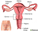
Vagina
The vagina is the female body part that connects the womb (uterus) and cervix to the outside of the body.

After the cervix is cleaned, the health care provider places a thin tube (catheter) through the cervix. Dye, called contrast, flows through this tube, filling the womb and fallopian tubes. X-rays are taken. The dye makes these areas easier to see on x-rays.
Cervix
The cervix is the lower end of the womb (uterus). It is at the top of the vagina. It is about 2. 5 to 3. 5 centimeters (1 to 1. 3 inches) long. Th...
x-rays
X-rays are a type of electromagnetic radiation, just like visible light. An x-ray machine sends individual x-ray waves through the body. The images...

How to Prepare for the Test
Your provider may give you antibiotics to take before and after the test. This helps prevent infections. You may also be given medicines to take the day of the procedure to help you relax.
The best time for this test is in the first half of the menstrual cycle. Doing it at this time enables the provider to see uterine cavity and tubes more clearly. It also reduces the risk for infection, and ensures that you are not pregnant.
Tell your provider if you have had an allergic reaction to contrast dye before.
You can eat and drink normally before the test.
How the Test will Feel
You may have some discomfort when the speculum is inserted into your vagina. This is similar to a pelvic exam with a Pap test.
Some women have cramps during or after the test, like those you may get during your period.
You may have some pain if the dye leaks out of the tubes, or if the tubes are blocked.
Why the Test is Performed
This test is done to check for blockages in your fallopian tubes or other problems in your womb and tubes. It is often done as part of an infertility exam. It may also be done after you have your tubes tied to confirm that the tubes are fully blocked after you have had a hysteroscopic tubal occlusion procedure to prevent pregnancy.
Normal Results
A normal result means everything looks normal. There are no defects.
Talk to your provider about the meaning of your specific test results.
What Abnormal Results Mean
Abnormal results may be due to:
- Developmental disorders of the structures of the uterus or fallopian tubes
- Scar tissue (adhesions) in the uterus or tubes
- Blockage of the fallopian tubes
- Presence of foreign bodies
- Tumors or polyps in the uterus
Risks
Risks may include:
- Allergic reaction to the contrast
- Endometrial infection (endometritis)
Endometritis
Endometritis is an inflammation or irritation of the lining of the uterus (the endometrium). It is not the same as endometriosis.
 ImageRead Article Now Book Mark Article
 - Fallopian tube infection (salpingitis)
Salpingitis
Pelvic inflammatory disease (PID) is an infection of a woman's womb (uterus), ovaries, or fallopian tubes.
 ImageRead Article Now Book Mark Article
 - Perforation of (poking a hole through) the uterus
Considerations
This test should not be performed if you have pelvic inflammatory disease (PID) or have unexplained vaginal bleeding.
Pelvic inflammatory disease
Pelvic inflammatory disease (PID) is an infection of a woman's womb (uterus), ovaries, or fallopian tubes.
Vaginal bleeding
This article discusses vaginal bleeding that occurs between a woman's monthly menstrual periods. Such bleeding may be called "intermenstrual bleedin...
After the test, tell your provider right away if you have any signs or symptoms of infection. These include foul-smelling vaginal discharge, pain, or fever. You may need to take antibiotics if this occurs.
Vaginal discharge
Vaginal discharge refers to secretions from the vagina. The discharge may be:Thick, pasty, or thinClear, cloudy, bloody, white, yellow, or greenOdor...
References
Fauser BCJM, Broekmans FJ. Female infertility: evaluation and management. In: Robertson RP, ed. DeFroot's Endocrinology. 8th ed. Philadelphia, PA: Elsevier; 2023:chap 123.
Fettinger S, Fanelli L. Hysterosalpingography and sonohysterography. In: Fowler GC, ed. Pfenninger and Fowler's Procedures for Primary Care. 4th ed. Philadelphia, PA: Elsevier; 2020:chap 134.
Lobo RA. Infertility: etiology, diagnostic evaluation, management, prognosis. In: Gershenson DM, Lentz GM, Valea FA, Lobo RA, eds. Comprehensive Gynecology. 8th ed. Philadelphia, PA: Elsevier; 2022:chap 40.
-
Uterus - illustration
The uterus is a hollow muscular organ located in the female pelvis between the bladder and rectum. The ovaries produce the eggs that travel through the fallopian tubes. Once the egg has left the ovary it can be fertilized and implant itself in the lining of the uterus. The main function of the uterus is to nourish the developing fetus prior to birth.
Uterus
illustration
Review Date: 3/31/2024
Reviewed By: LaQuita Martinez, MD, Department of Obstetrics and Gynecology, Emory Johns Creek Hospital, Alpharetta, GA. Also reviewed by David C. Dugdale, MD, Medical Director, Brenda Conaway, Editorial Director, and the A.D.A.M. Editorial team.
 All rights reserved.