

Abnormally dark or light skin
Hyperpigmentation; Hypopigmentation; Skin - abnormally light or darkAbnormally dark or light skin is skin that has turned darker or lighter than normal.
Considerations
Normal skin contains cells called melanocytes. These cells produce melanin, the substance that gives skin its color.
Melanin
Melanin is a natural substance that gives color (pigment) to: HairSkinThe iris of the eye It is produced by cells in the skin called melanocytes. Mel...

Skin with too much melanin is called hyperpigmented skin.
Skin with too little melanin is called hypopigmented. Skin with no melanin at all is called depigmented.
Pale skin areas are due to too little melanin or underactive melanocytes. Darker areas of skin (or an area that tans more easily) occurs when you have more melanin or overactive melanocytes.
Pale
Paleness is an abnormal loss of color from normal skin or mucous membranes.
Read Article Now Book Mark ArticleBronzing of the skin may sometimes be mistaken for a suntan. This skin discoloration often develops slowly, starting at the elbows, knuckles, and knees and spreading from there. Bronzing may also be seen on the soles of the feet and the palms of the hands. The bronze color can range from light to dark (in fair-skinned people) with the degree of darkness due to the underlying cause.
Causes
Causes of hyperpigmentation include:
- Skin inflammation (post-inflammatory hyperpigmentation)
- Use of certain drugs (such as minocycline, certain cancer chemotherapies, and birth control pills)
- Hormone system diseases such as Addison disease
Addison disease
Addison disease is a disorder that causes the adrenal glands to not produce enough hormones.
 ImageRead Article Now Book Mark Article
 - Hemochromatosis (iron overload)
- Sun exposure
- Pregnancy (melasma, or mask of pregnancy)
- Certain birthmarks
- Skin condition called acanthosis nigricans
Causes of hypopigmentation include:
- Skin inflammation (post-inflammatory hypopigmentation)
- Certain fungal infections (such as tinea versicolor)
Tinea versicolor
Tinea versicolor is a common fungal infection of the outer layer of the skin.
 ImageRead Article Now Book Mark Article
 -
Pityriasis alba
Pityriasis alba
Pityriasis alba is a common skin disorder that causes patches of light-colored (hypopigmented) areas.
Read Article Now Book Mark Article -
Vitiligo
Vitiligo
Vitiligo is a skin condition in which there is a loss of color (pigment) from areas of skin. This results in uneven white patches that have no pigme...
 ImageRead Article Now Book Mark Article
 - Certain medicines
- Skin condition called idiopathic guttate hypomelanosis in sun exposed areas such as the arms
- Certain birthmarks
Home Care
Over-the-counter and prescription creams are available for lightening the skin. Hydroquinone combined with tretinoin is an effective combination. If you use these creams, follow instructions carefully, and don't use one for more than 3 weeks at a time. Darker skin requires greater care when using these preparations. Cosmetics may also help mask a discoloration.
Avoid too much sun exposure. Always use sunscreen with an SPF of 30 or higher.
Abnormally dark skin may continue even after treatment.
When to Contact a Medical Professional
Call your health care provider for an appointment if you have:
- Skin discoloration that causes significant concern
- Persistent, unexplained darkening or lightening of the skin
- Any skin sore or lesion that changes shape, size, or color may be a sign of skin cancer
What to Expect at Your Office Visit
Your provider will perform a physical exam and ask about your symptoms, including:
- When did the discoloration develop?
- Did it develop suddenly?
- Is it getting worse? How fast?
- Has it spread to other parts of the body?
- What medicines do you take?
- Has anyone else in your family had a similar problem?
- How often are you in the sun? Do you use a sun lamp or go to tanning salons?
- What is your diet like?
- What other symptoms do you have? For example, are there any rashes or skin lesions?
Skin lesions
Rashes involve changes in the color, feeling or texture of your skin.
 ImageRead Article Now Book Mark Article
Tests that may be done include:
- Adrenocorticotrophin hormone stimulation test
-
Skin biopsy
Skin biopsy
A skin lesion biopsy is when a small amount of skin is removed so it can be examined under a microscope. The skin is tested to look for skin conditi...
 ImageRead Article Now Book Mark Article
 -
Thyroid function studies
Thyroid function studies
Thyroid function tests are used to check whether your thyroid is working normally. The most common thyroid function tests are:Free T4 (the main thyro...
 ImageRead Article Now Book Mark Article
 -
Wood lamp test
Wood lamp test
A Wood lamp examination is a test that uses ultraviolet (UV) light to look at the skin closely.
 ImageRead Article Now Book Mark Article
 -
KOH test
KOH test
The skin lesion KOH exam is a test to diagnose a fungal infection of the skin.
 ImageRead Article Now Book Mark Article
Your provider may recommend creams, ointments, surgery, or phototherapy, depending on the type of skin condition you have. Bleaching creams can help lighten dark areas of skin.
Some skin color changes may return to normal without treatment.
References
Chang MW. Disorders of hyperpigmentation. In: Bolognia JL, Schaffer JV, Cerroni L, eds. Dermatology. 4th ed. Philadelphia, PA: Elsevier; 2018:chap 67.
Passeron T, Ortonne JP. Vitiligo and other disorders of hypopigmentation. In: Bolognia JL, Schaffer JV, Cerroni L, eds. Dermatology. 4th ed. Philadelphia, PA: Elsevier; 2018:chap 66.
-
Vitiligo - drug induced - illustration
The white spots on this person's face have resulted from drug-induced vitiligo. Loss of melanin, the primary skin pigment, occasionally occurs as a result of medicines, as is the case with this individual. The typical vitiligo lesion is flat and depigmented, but maintains the normal skin texture.
Vitiligo - drug induced
illustration
-
Vitiligo on the face - illustration
This is a picture of vitiligo on the face. Complete loss of melanin, the primary skin pigment, occurs for unknown reasons. The resulting lesions are white in comparison to the surrounding skin. Vitiligo may occur in the same areas on both sides of the face -- symmetrically -- or it may be patchy -- asymmetrical. The typical vitiligo lesion is flat and depigmented, but maintains the normal skin texture. The dark areas around the eyes are this person's normal skin color.
Vitiligo on the face
illustration
-
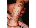
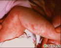
Incontinentia pigmenti on the leg - illustration
Incontinentia pigmenti produces darkly-pigmented swirling marks on the skin. It occurs more frequently in females. The skin lesions are divided into three stages blisters (vesicles and bullae) are present at birth or within the first 6 to 7 weeks, followed by a rough wart-like (verrucous) stage, and lastly, swirled and bizarre patterns of dark pigmentation (hyperpigmentation) appear.
Incontinentia pigmenti on the leg
illustration
-
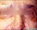
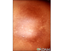
Hyperpigmentation 2 - illustration
Hyperpigmentation refers to skin that has turned darker than normal where the change that has occurred is unrelated to sun exposure. Cells called melanocytes located in the skin, produce melanin. Melanin gives the skin its color. In certain conditions melanocytes can become abnormal and cause an excessive amount of darkening in the color of the skin.
Hyperpigmentation 2
illustration
-
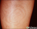
Post-inflammatory hyperpigmentation - calf - illustration
Hyperpigmented concentric rings over the tibia are secondary to prior inflammation. Residual hemosiderin from broken down red blood cells in macrophages, left behind after inflammation pigments the skin.
Post-inflammatory hyperpigmentation - calf
illustration
-
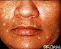
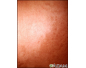
Hyperpigmentation w/malignancy - illustration
Generalized hyperpigmentation, in addition to localized areas of even deeper pigmentation is sometimes found in patients with malignancy. Etiology of these pigmentary changes is varied.
Hyperpigmentation w/malignancy
illustration
-
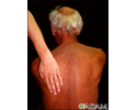
Post-inflammatory hyperpigmentation 2 - illustration
Post-inflammatory hyperpigmentation is seen here in a mottled pattern, over the posterior shoulder. Hemosiderin, left behind by degraded RBC's, creates the discoloration.
Post-inflammatory hyperpigmentation 2
illustration
-
Vitiligo - drug induced - illustration
The white spots on this person's face have resulted from drug-induced vitiligo. Loss of melanin, the primary skin pigment, occasionally occurs as a result of medicines, as is the case with this individual. The typical vitiligo lesion is flat and depigmented, but maintains the normal skin texture.
Vitiligo - drug induced
illustration
-
Vitiligo on the face - illustration
This is a picture of vitiligo on the face. Complete loss of melanin, the primary skin pigment, occurs for unknown reasons. The resulting lesions are white in comparison to the surrounding skin. Vitiligo may occur in the same areas on both sides of the face -- symmetrically -- or it may be patchy -- asymmetrical. The typical vitiligo lesion is flat and depigmented, but maintains the normal skin texture. The dark areas around the eyes are this person's normal skin color.
Vitiligo on the face
illustration
-
Incontinentia pigmenti on the leg - illustration
Incontinentia pigmenti produces darkly-pigmented swirling marks on the skin. It occurs more frequently in females. The skin lesions are divided into three stages blisters (vesicles and bullae) are present at birth or within the first 6 to 7 weeks, followed by a rough wart-like (verrucous) stage, and lastly, swirled and bizarre patterns of dark pigmentation (hyperpigmentation) appear.
Incontinentia pigmenti on the leg
illustration
-
Hyperpigmentation 2 - illustration
Hyperpigmentation refers to skin that has turned darker than normal where the change that has occurred is unrelated to sun exposure. Cells called melanocytes located in the skin, produce melanin. Melanin gives the skin its color. In certain conditions melanocytes can become abnormal and cause an excessive amount of darkening in the color of the skin.
Hyperpigmentation 2
illustration
-
Post-inflammatory hyperpigmentation - calf - illustration
Hyperpigmented concentric rings over the tibia are secondary to prior inflammation. Residual hemosiderin from broken down red blood cells in macrophages, left behind after inflammation pigments the skin.
Post-inflammatory hyperpigmentation - calf
illustration
-
Hyperpigmentation w/malignancy - illustration
Generalized hyperpigmentation, in addition to localized areas of even deeper pigmentation is sometimes found in patients with malignancy. Etiology of these pigmentary changes is varied.
Hyperpigmentation w/malignancy
illustration
-
Post-inflammatory hyperpigmentation 2 - illustration
Post-inflammatory hyperpigmentation is seen here in a mottled pattern, over the posterior shoulder. Hemosiderin, left behind by degraded RBC's, creates the discoloration.
Post-inflammatory hyperpigmentation 2
illustration
-
Psoriasis-In-Depth
(In-Depth)
-
Diabetes - type 2 - InDepth
(In-Depth)
-
Cirrhosis - InDepth
(In-Depth)
-
Menstrual disorders - InDepth
(In-Depth)
-
Hepatitis - InDepth
(In-Depth)
-
Gallstones and gallbladder disease - InDepth
(In-Depth)
Review Date: 6/7/2023
Reviewed By: Elika Hoss, MD, Assistant Professor of Dermatology, Mayo Clinic, Scottsdale, AZ. Also reviewed by David C. Dugdale, MD, Medical Director, Brenda Conaway, Editorial Director, and the A.D.A.M. Editorial team.
 All rights reserved.