Head and face reconstruction
Craniofacial reconstruction; Orbital-craniofacial surgery; Facial reconstructionHead and face reconstruction is surgery to repair or reshape deformities of the head and face (craniofacial).
Description
How surgery for head and face deformities (craniofacial reconstruction) is done depends on the type and severity of deformity, and the person's condition. The medical term for this surgery is craniofacial reconstruction.
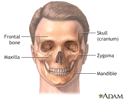
Surgical repairs involve the skull (cranium), brain, nerves, eyes, and the bones and skin of the face. That is why sometimes a plastic surgeon (for skin and face) and a neurosurgeon (brain and nerves) work together. Head and neck surgeons also perform craniofacial reconstruction operations.
The surgery is done while you are deep asleep and pain-free (under general anesthesia). The surgery may take 4 to 12 hours or more. Some of the bones of the face are cut and moved. During the surgery, tissues are moved and blood vessels and nerves are reconnected using microscopic surgery techniques.
General anesthesia
General anesthesia is treatment with certain medicines that puts you into a deep sleep-like state so you do not feel pain during surgery. After you ...
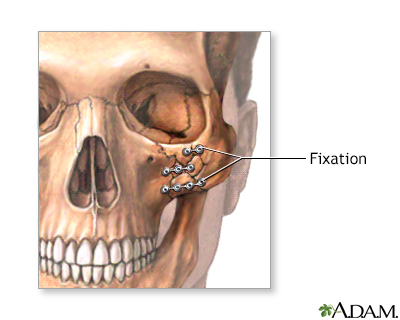
Read Article Now Book Mark ArticlePieces of bone (bone grafts) may be taken from the pelvis, ribs, or skull to fill in spaces where bones of the face and head were moved. Small screws and plates made of titanium or a fixation device made of absorbable material may be used to hold the bones in place. Implants may also be used to help reshape the skull. The jaws may be wired together to hold the new bone positions in place. To cover the holes, flaps of tissue may be taken from the hand, buttocks, chest wall, or thigh.
Bone grafts
A bone graft is surgery to place new bone or bone substitutes into spaces around a broken bone or bone defects to stimulate healing.

Sometimes the surgery causes swelling of the face, mouth, or neck, which may last for weeks to months. This can block the airway. For this reason, you may need to have a temporary tracheostomy. This is a small hole that is made in your neck through which a tube (endotracheal tube) is placed in the airway (trachea). This allows you to breathe when your face and upper airway are swollen.
Tracheostomy
A tracheostomy is a surgical procedure to create an opening through the neck into the trachea (windpipe). A tube is most often placed through this o...

Why the Procedure Is Performed
Craniofacial reconstruction may be done if there are:
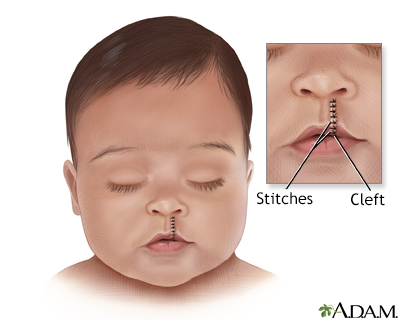
- Birth defects and deformities from conditions such as cleft lip or palate, craniosynostosis, Apert syndrome
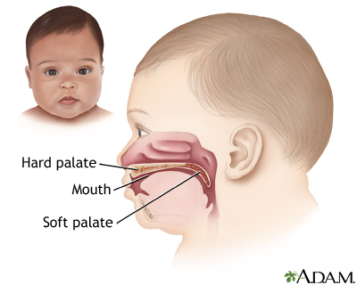
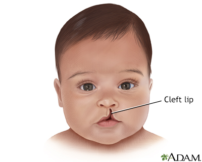
Cleft lip or palate
Cleft lip and palate are birth defects that affect the upper lip and the roof of the mouth.
 ImageRead Article Now Book Mark Article
Craniosynostosis
Craniosynostosis is a birth defect in which one or more sutures on a baby's head closes earlier than usual. The skull of an infant or young child is ...
 ImageRead Article Now Book Mark Article
Apert syndrome
Apert syndrome is a genetic disease in which the seams between the skull bones close earlier than normal. This affects the shape of the head and fac...
 ImageRead Article Now Book Mark Article
 - Deformities caused by surgery done to treat tumors
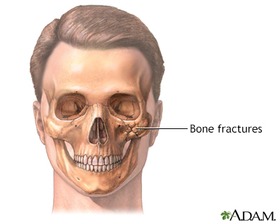
- Injuries to the head, face, or jaw
- Tumors
Risks
Risks of anesthesia and surgery in general are:
- Problems breathing
- Reactions to medicines
- Bleeding, blood clots, infection
Risks of surgery of the head and face are:
- Nerve (cranial nerve dysfunction) or brain damage
- Need for follow-up surgery, especially in growing children
- Partial or total loss of bone grafts
- Permanent scarring
These complications are more common in people who:
- Smoke
- Have poor nutrition
- Have other medical conditions, such as lupus
Lupus
Systemic lupus erythematosus (SLE) is an autoimmune disease. In this disease, the immune system of the body mistakenly attacks healthy tissue. It c...
 ImageRead Article Now Book Mark Article
 - Have poor blood circulation
- Have past nerve damage
After the Procedure
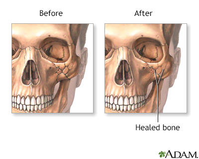
You may spend the first 2 days after surgery in the intensive care unit. If you do not have a complication, you will be able to leave the hospital usually within 1 week. More limited procedures may require shorter or no hospital stay. Complete healing may take 6 weeks or more. Swelling will improve over the following months.
Outlook (Prognosis)
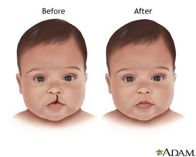
A much more normal appearance can be expected after surgery. Some people need to have follow-up procedures during the next 1 to 4 years.
It is important not to play contact sports for 2 to 6 months after surgery.
People who have had a serious injury often need to work through the emotional issues of the trauma and the change in their appearance. Children and adults who have had a serious injury may have post-traumatic stress disorder, depression, and anxiety disorders. Talking to a mental health professional or joining a support group can be helpful.
Parents of children with deformities of the face often feel guilty or ashamed, especially when the deformities are due to a genetic condition. As children grow and become aware of their appearance, emotional symptoms may develop or get worse.
References
Baker SR. Reconstruction of facial defects. In: Flint PW, Francis HW, Haughey BH, et al, eds. Cummings Otolaryngology: Head & Neck Surgery. 7th ed. Philadelphia, PA: Elsevier; 2021:chap 21.
Padilla PL, Khoo KH, Ho T, Cole EL, Sirvent RZ, Philips LG. Plastic surgery. In: Townsend CM Jr, Beauchamp RD, Evers BM, Mattox KL, eds. Sabiston Textbook of Surgery: The Biological Basis of Modern Surgical Practice. 21st ed. St Louis, MO: Elsevier; 2022:chap 69.
-
Skull - illustration
The skull is anterior to the spinal column and is the bony structure that encases the brain. Its purpose is to protect the brain and allow attachments for the facial muscles. The two regions of the skull are the cranial and facial region. The cranial portion is the part of the skull that directly houses the brain and the facial portion includes the rest of the bones of the skull.
Skull
illustration
-
Skull - illustration
The skull is the bony structure of the head and face. The cranium surrounds the brain with the temporal, frontal, parietal and occipital bones. The maxilla, or upper jaw, and the mandible, or lower jaw, support the facial features of nose, mouth and eyes.
Skull
illustration
-
Cleft lip repair - series
Presentation
-
Craniofacial reconstruction - series
Presentation
-
Skull - illustration
The skull is anterior to the spinal column and is the bony structure that encases the brain. Its purpose is to protect the brain and allow attachments for the facial muscles. The two regions of the skull are the cranial and facial region. The cranial portion is the part of the skull that directly houses the brain and the facial portion includes the rest of the bones of the skull.
Skull
illustration
-
Skull - illustration
The skull is the bony structure of the head and face. The cranium surrounds the brain with the temporal, frontal, parietal and occipital bones. The maxilla, or upper jaw, and the mandible, or lower jaw, support the facial features of nose, mouth and eyes.
Skull
illustration
-
Cleft lip repair - series
Presentation
-
Craniofacial reconstruction - series
Presentation
Review Date: 5/26/2023
Reviewed By: Tang Ho, MD, Associate Professor, Division of Facial Plastic and Reconstructive Surgery, Department of Otolaryngology - Head and Neck Surgery, The University of Texas Medical School at Houston, Houston, TX. Also reviewed by David C. Dugdale, MD, Medical Director, Brenda Conaway, Editorial Director, and the A.D.A.M. Editorial team.


