Heart valve surgery
Valve replacement; Valve repair; Heart valve prosthesis; Mechanical valves; Prosthetic valvesHeart valve surgery is used to repair or replace diseased heart valves.
Blood that flows between different chambers of your heart must flow through a heart valve. Blood that flows out of your heart into large arteries must also flow through a heart valve.
These valves open up enough so that blood can flow through. They then close, keeping blood from flowing backward.
There are 4 valves in your heart:
- Aortic valve
- Mitral valve
- Tricuspid valve
- Pulmonic valve
The aortic valve is the most common valve to be replaced. The mitral valve is the most common valve to be repaired. Only rarely is the tricuspid valve or the pulmonic valve repaired or replaced.
Description
Before your surgery, you will receive general anesthesia. You will be asleep and unable to feel pain.
General anesthesia
General anesthesia is treatment with certain medicines that puts you into a deep sleep-like state so you do not feel pain during surgery. After you ...
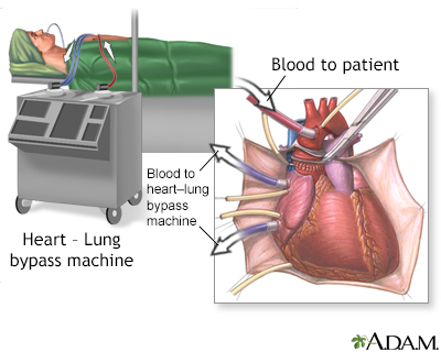
Read Article Now Book Mark ArticleIn open heart surgery, the heart surgeon makes a large surgical cut in your breastbone to reach the heart and aorta. You are connected to a heart-lung bypass machine. Your heart is stopped while you are connected to this machine. This machine does the work of your heart, providing oxygen and removing carbon dioxide.
Minimally invasive valve surgery is done through much smaller cuts than open surgery, or through a catheter inserted through the skin. Several different techniques are used:
- Percutaneous surgery (through the skin)
- Robot-assisted surgery
Robot-assisted surgery
Robotic surgery is a method to perform surgery using very small tools attached to a robotic arm. The surgeon controls the robotic arm with a compute...
Read Article Now Book Mark Article
If your surgeon can repair your mitral valve, you may have:
- Ring annuloplasty. The surgeon repairs the ring-like part around the valve by sewing a ring of plastic, cloth, or tissue around the valve.
- Valve repair. The surgeon trims, shapes, or rebuilds one or more of the leaflets of the valve. The leaflets are flaps that open and close the valve. Valve repair is best for the mitral and tricuspid valves. The aortic valve is usually not repaired.
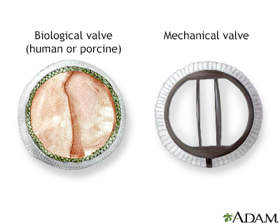
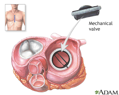
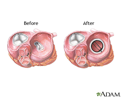
If your valve is too damaged, you will need a new valve. This is called valve replacement surgery. Your surgeon will remove your valve and put a new one in place. The main types of new valves are:
- Mechanical -- made of man-made materials, such as metal (stainless steel or titanium) or ceramic. These valves last the longest, but you will need to take blood-thinning medicine, such as warfarin (Coumadin) or aspirin, for the rest of your life.
- Biological -- made of human or animal tissue. These valves last 12 to 15 years, but you may not need to take blood thinners for life.
In some cases, surgeons can use your own pulmonic valve to replace the damaged aortic valve. The pulmonic valve is then replaced with an artificial valve (this is called the Ross Procedure). This procedure may be useful for people who do not want to take blood thinners for the rest of their life. However, the new aortic valve does not last very long and may need to be replaced again by either a mechanical or a biologic valve.
Related topics include:
- Aortic valve surgery - minimally invasive
Aortic valve surgery - minimally invasi...
Blood flows out of your heart and into a large blood vessel called the aorta. The aortic valve separates the heart and aorta. The aortic valve open...
 ImageRead Article Now Book Mark Article
 - Aortic valve surgery - open
Aortic valve surgery - open
Blood flows out of your heart and into a large blood vessel called the aorta. The aortic valve separates the heart and aorta. The aortic valve open...
 ImageRead Article Now Book Mark Article
 - Mitral valve surgery - minimally invasive
Mitral valve surgery - minimally invasi...
Mitral valve surgery is surgery to either repair or replace the mitral valve in your heart. Blood flows from the lungs and enters a pumping chamber o...
 ImageRead Article Now Book Mark Article
 - Mitral valve surgery - open
Mitral valve surgery - open
Mitral valve surgery is used to repair or replace the mitral valve in your heart. Blood flows between the different chambers in the heart through val...
Read Article Now Book Mark Article
Why the Procedure Is Performed
You may need surgery if your valve does not work properly.
- A valve that does not close all the way will allow blood to leak backwards. This is called regurgitation.
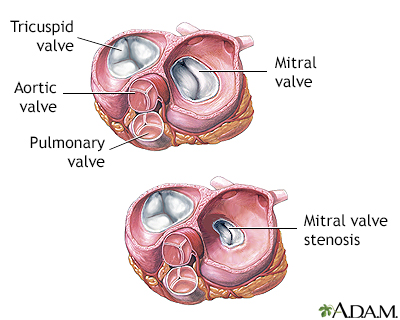
- A valve that does not open fully will limit forward blood flow. This is called stenosis.
You may need heart valve surgery for these reasons:
- Defects in your heart valve are causing major heart symptoms, such as chest pain (angina), shortness of breath, fainting spells (syncope), or heart failure.
Angina
Angina is a type of chest discomfort or pain due to poor blood flow through the blood vessels (coronary arteries) of the heart muscle (myocardium). ...
 ImageRead Article Now Book Mark Article
Fainting spells
Fainting is a brief loss of consciousness due to a drop in blood flow to the brain. The episode most often lasts less than a couple of minutes and y...
Read Article Now Book Mark Article - Tests show that the changes in your heart valve are beginning to seriously affect your heart function.
- Your doctor wants to replace or repair your heart valve at the same time as you are having open heart surgery for another reason, such as a coronary artery bypass graft surgery.
- Your heart valve has been damaged by infection (endocarditis).
Endocarditis
Endocarditis is inflammation of the inside lining of the heart chambers and heart valves (endocardium). It is most often caused by a bacterial or, r...
 ImageRead Article Now Book Mark Article
 - You have received a new heart valve in the past and it is not working well, or you have other problems such as blood clots, infection, or bleeding.
Some of the heart valve problems treated with surgery are:
- Aortic insufficiency
- Aortic stenosis
- Congenital heart valve disease
- Mitral regurgitation - acute
- Mitral regurgitation - chronic
- Mitral stenosis
- Mitral valve prolapse
- Pulmonary valve stenosis
- Tricuspid regurgitation
- Tricuspid valve stenosis
Risks
The risks of having heart surgery include:
- Death
- Heart attack
- Heart failure
- Bleeding requiring reoperation
- Rupture of the heart
- Irregular heartbeat (arrhythmia)
Arrhythmia
An arrhythmia is a disorder of the heart rate (pulse) or heart rhythm. The heart can beat too fast (tachycardia), too slow (bradycardia), or irregul...
 ImageRead Article Now Book Mark Article
 - Kidney failure
- Post-pericardiotomy syndrome -- low fever and chest pain that can last for up to 6 months
- Stroke or other temporary or permanent brain injury
- Infection
- Problems with the breastbone healing
- Temporary confusion after surgery due to the heart-lung machine
It is very important to take steps to prevent valve infections. You may need to take antibiotics before dental work and other invasive procedures.
Before the Procedure
Your preparation for the procedure will depend on the type of valve surgery you are having:
- Aortic valve surgery - minimally invasive
- Aortic valve surgery - open
- Mitral valve surgery - minimally invasive
- Mitral valve surgery - open
After the Procedure
Your recovery after the procedure will depend on the type of valve surgery you are having:
- Aortic valve surgery - minimally invasive
- Aortic valve surgery - open
- Mitral valve surgery - minimally invasive
- Mitral valve surgery - open
The average hospital stay is 5 to 7 days. The nurse will tell you how to care for yourself at home. Complete recovery will take a few weeks to several months, depending on your health before surgery.
How to care for yourself at home
Heart valve surgery is used to repair or replace diseased heart valves. Your surgery may have been done through a large incision (cut) in the middle...
Read Article Now Book Mark ArticleOutlook (Prognosis)
The success rate of heart valve surgery is high. The operation can relieve your symptoms and prolong your life.
Mechanical heart valves do not often fail. However, blood clots can develop on these valves. If a blood clot forms, you may have a stroke. Bleeding can occur, but this is rare. Tissue valves last an average of 12 to 15 years, depending on the type of valve. Long-term use of blood thinning medicine is most often not needed with tissue valves.
There is always a risk for infection. Talk to your health care provider and surgeon before having any type of medical procedure.
The clicking of mechanical heart valves may be heard in the chest. This is normal.
References
Carabello BA, Kodali S. Valvular heart disease. In: Goldman L, Cooney KA, eds. Goldman-Cecil Medicine. 27th ed. Philadelphia, PA: Elsevier; 2024:chap 60.
Herrmann HC, Reardon MJ. Transcatheter therapies for mitral and tricuspid valvular heart disease. In: Libby P, Bonow RO, Mann DL, Tomaselli, GF, Bhatt DL, Solomon SD, eds. Braunwald's Heart Disease: A Textbook of Cardiovascular Medicine. 12th ed. Philadelphia, PA: Elsevier; 2022:chap 78.
Leon MB, Mack MJ. Transcatheter aortic valve replacement. In: Libby P, Bonow RO, Mann DL, Tomaselli, GF, Bhatt DL, Solomon SD, eds. Braunwald's Heart Disease: A Textbook of Cardiovascular Medicine. 12th ed. Philadelphia, PA: Elsevier; 2022:chap 74.
Pellikka PA, Nkomo VT. Tricuspid, pulmonic, and multivalvular disease. In: Libby P, Bonow RO, Mann DL, Tomaselli, GF, Bhatt DL, Solomon SD, eds. Braunwald's Heart Disease: A Textbook of Cardiovascular Medicine. 12th ed. Philadelphia, PA: Elsevier; 2022:chap 77.
Otto CM, Nishimura RA, Bonow RO, et al. 2020 ACC/AHA guideline for the management of patients with valvular heart disease: executive summary: a report of the American College of Cardiology/American Heart Association Joint Committee on clinical practice guidelines. Circulation. 2021;143(5):e35-e71. PMID: 33332149 pubmed.ncbi.nlm.nih.gov/33332149/.
Rosengart TK, Aberle CM, Ryan C. Acquired heart disease: valvular. In: Townsend CM Jr, Beauchamp RD, Evers BM, Mattox KL, eds. Sabiston Textbook of Surgery. 21st ed. Philadelphia, PA: Elsevier; 2022:chap 61.
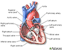
Heart - section through the middle - illustration
The interior of the heart is composed of valves, chambers, and associated vessels.
Heart - section through the middle
illustration
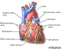
Heart - front view - illustration
The external structures of the heart include the ventricles, atria, arteries and veins. Arteries carry blood away from the heart while veins carry blood into the heart. The vessels colored blue indicate the transport of blood with relatively low content of oxygen and high content of carbon dioxide. The vessels colored red indicate the transport of blood with relatively high content of oxygen and low content of carbon dioxide.
Heart - front view
illustration
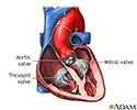
Heart valves - anterior view - illustration
There are four valves located in the heart. Each valve either consists of two or three folds of thin tissue. When closed, the valve prevents blood from flowing backwards to its previous location. When open the valve allows blood to flow freely. Valve problems can occur because of congenital abnormalities, infection, or other causes.
Heart valves - anterior view
illustration
Heart valves - superior view - illustration
There are four valves located in the heart. Each valve either consists of two or three folds of thin tissue. When closed, the valve prevents blood from flowing backwards to its previous location. When open the valve allows blood to flow freely. Valve problems can occur because of congenital abnormalities, infection, or other causes.
Heart valves - superior view
illustration
Heart valve surgery - Series
Presentation
- Heart failure - InDepth(In-Depth)
- Heart attack and acute coronary syndrome - InDepth(In-Depth)
Review Date: 5/13/2024
Reviewed By: Mary C. Mancini, MD, PhD, Cardiothoracic Surgeon, Shreveport, LA. Review provided by VeriMed Healthcare Network. Also reviewed by David C. Dugdale, MD, Medical Director, Brenda Conaway, Editorial Director, and the A.D.A.M. Editorial team.

