

Congenital heart defect - corrective surgery
Congenital heart surgery; Patent ductus arteriosus ligation; Hypoplastic left heart repair; Tetralogy of Fallot repair; Coarctation of the aorta repair; Atrial septal defect repair; Ventricular septal defect repair; Truncus arteriosus repair; Total anomalous pulmonary artery correction; Transposition of great vessels repair; Tricuspid atresia repair; VSD repair; ASD repairCongenital heart defect corrective surgery fixes or treats a heart defect that a child is born with. A baby born with one or more heart defects has congenital heart disease. Surgery is needed if the defect could harm the child's long-term health or well-being. Sometimes, congenital heart disease is not detected until adulthood.
Congenital heart disease
Congenital heart disease (CHD) is a problem with the heart's structure and function that is present at birth.

Description
There are many types of pediatric heart surgery.
Pediatric heart surgery
Heart surgery in children is done to repair heart defects a child is born with (congenital heart defects) and heart diseases a child gets after birth...

Patent ductus arteriosus (PDA) ligation:
Patent ductus arteriosus
Patent ductus arteriosus (PDA) is a condition in which the ductus arteriosus does not close. The word "patent" means open. The ductus arteriosus is ...
- Before birth, the baby has a blood vessel that runs between the aorta (the main artery to the body) and the pulmonary artery (the main artery to the lungs), called the ductus arteriosus. This small vessel most often closes shortly after birth when the baby starts to breathe on their own. If it does not close. It is called a patent ductus arteriosus. This could cause problems later in life.
- In most cases, the cardiologist will close off the opening using medicine. If this does not work, then other techniques are used.
- Sometimes the PDA can be closed with a procedure that does not involve surgery. The procedure is most often done in a laboratory that uses x-rays. In this procedure, the surgeon makes a small cut in the groin. A wire and tube called a catheter is inserted into an artery in the leg and passed up to the heart. Then, a small metal coil or another device is passed through the catheter into the infant's ductus arteriosus artery. The coil or other device blocks the blood flow, and this corrects the problem.
- Another method is to make a small surgical cut on the left side of the chest. The surgeon finds the PDA and then ties off or clips the ductus arteriosus, or divides and cuts it. Tying off the ductus arteriosus is called ligation. This procedure may be done in the neonatal intensive care unit (NICU).
Coarctation of the aorta repair:
Coarctation of the aorta
The aorta is a larger artery that carries blood from the heart to the vessels that supply the rest of the body with blood. If part of the aorta is n...

- Coarctation of the aorta occurs when a part of the aorta has a very narrow section. The shape looks like an hourglass timer. The narrowing makes it difficult for blood to get through to the lower extremities. Over time, it can lead to problems such as extremely high blood pressure.
- To repair this defect, a cut is most often made on the left side of the chest, between the ribs. There are several ways to repair coarctation of the aorta.
- The most common way to repair it is to cut the narrow section and make it bigger with a patch made of GORE-TEX, a man-made (synthetic) material.
- Another way to repair this problem is to remove the narrow section of the aorta and stitch the remaining ends together. This can most often be done in older children.
- A third way to repair this problem is called a subclavian flap. First, a cut is made in the narrow part of the aorta. Then, a patch is taken from the left subclavian artery (the artery to the arm) to enlarge the narrow section of the aorta.
- A fourth way to repair the problem is to connect a tube to the normal sections of the aorta, on either side of the narrow section. Blood flows through the tube and bypasses the narrow section.
- A newer method does not require surgery. A small wire is placed through an artery in the groin and up to the aorta. A small balloon is then opened up in the narrow area of the aorta. A stent or small tube is left there to help keep the aorta open. The procedure is done in a laboratory with x-rays. This procedure is often used when the coarctation reoccurs after it has been fixed.
Atrial septal defect (ASD) repair:
Atrial septal defect
Atrial septal defect (ASD) is a heart defect that is present at birth (congenital). As a baby develops in the womb, a wall (septum) forms that divide...

- The atrial septum is the wall between the left and right atria (upper chambers) of the heart. A hole in that wall is called an ASD. In the presence of this defect, blood with and without oxygen can be mixed up over time, causing medical problems and arrhythmias.
- Sometimes, an ASD can be closed without open-heart surgery. First, the surgeon makes a tiny cut in the groin. Then the surgeon inserts a wire into a blood vessel that goes to the heart. Next, two small umbrella-shaped "clamshell" devices are placed on the right and left sides of the septum. These two devices are attached to each other. This closes the hole in the heart. Not all medical centers do this procedure.
- Open-heart surgery may also be done to repair ASD. In this operation, the septum can be closed using stitches. Another way to cover the hole is with a patch.
Ventricular septal defect (VSD) repair:
Ventricular septal defect
Ventricular septal defect is a hole in the wall that separates the right and left ventricles of the heart. Ventricular septal defect is one of the m...
- The ventricular septum is the wall between the left and right ventricles (lower chambers) of the heart. A hole in the ventricular septum is called a VSD. This hole lets blood with oxygen mix with used blood returning to the lungs. Over time, irregular heartbeats and other heart problems can happen.
- By age 1, most small VSDs close on their own. However, those VSDs that do stay open after this age may need to be closed with surgery.
- Larger VSDs and small ones in certain parts of the ventricular septum, or ones that cause heart failure or endocarditis, (inflammation) need open-heart surgery. The hole in the septum is most often closed with a patch.
- Some septal defects can be closed without surgery. The procedure involves passing a small wire into the heart and placing a small device to close the defect.
Tetralogy of Fallot repair:
Tetralogy of Fallot
Tetralogy of Fallot is a type of congenital heart defect. Congenital means that it is present at birth.
- Tetralogy of Fallot is a heart defect that usually includes four defects in the heart and causes the baby to turn a bluish color (cyanosis).
Cyanosis
A bluish color to the skin or mucous membrane is usually due to a lack of oxygen in the blood. The medical term is cyanosis.
 ImageRead Article Now Book Mark Article
 - Open-heart surgery is needed, and it is often done when the child is between 6 months and 2 years old.
- The surgery involves closing the VSD with a patch and opening the thickened muscle (stenosis). A patch is placed on the right ventricle and main pulmonary artery to improve blood flow to the lungs.
- The child may have a shunt procedure done first. A shunt moves blood from one area to another. This is done if the open-heart surgery needs to be delayed because the child is too sick to go through surgery.
- During a shunt procedure, the surgeon makes a surgical cut in the left side of the chest.
- Once the child is older, the shunt is closed and the main repair in the heart is performed.
Transposition of the great vessels repair:
Transposition of the great vessels
Transposition of the great arteries (TGA) is a heart defect that occurs from birth (congenital). The two major arteries that carry blood away from t...
- In a normal heart, the aorta comes from the left side of the heart, and the pulmonary artery comes from the right side. In transposition of the great vessels, these arteries come from the opposite sides of the heart. The child may also have other birth defects.
- Correcting transposition of the great vessels requires open-heart surgery. If possible, this surgery is done shortly after birth.
- The most common repair is called an arterial switch. The aorta and pulmonary artery are divided. The pulmonary artery is connected to the right ventricle, where it belongs. Then, the aorta and coronary arteries are connected to the left ventricle, where they belong.
Truncus arteriosus repair:
Truncus arteriosus
Truncus arteriosus is a rare type of heart disease in which a single blood vessel (truncus arteriosus) comes out of the right and left ventricles, in...
- Truncus arteriosus is a rare condition that occurs when the aorta, coronary arteries, and pulmonary artery all come out of one common trunk. The disorder may be very simple, or very complex. In all cases, it requires open-heart surgery to repair the defect.
- Repair is usually done in the first few days or weeks of the infant's life. The pulmonary arteries are separated from the aortic trunk, and any defects are patched. Usually, children also have a ventricular septal defect, and that is also closed. A connection is then placed between the right ventricle and the pulmonary arteries.
- Most children need one or two more surgeries as they grow.
Tricuspid atresia repair:
Tricuspid atresia
Tricuspid atresia is a type of heart disease that is present at birth (congenital heart disease), in which the tricuspid heart valve is missing or ab...
- The tricuspid valve is between the upper and lower chambers on the right side of the heart. Tricuspid atresia occurs when this valve is deformed, narrow, or missing.
- Babies born with tricuspid atresia are blue because they cannot get blood to the lungs to pick up oxygen.
- To get to the lungs, blood must cross an atrial septal defect (ASD), ventricular septal defect (VSD), or a patent ductus arteriosus (PDA). (These conditions are described above.) This condition severely restricts blood flow to the lungs.
- Soon after birth, the baby may be given a medicine called prostaglandin E. This medicine will help keep the patent ductus arteriosus open so that blood can continue to flow to the lungs. However, this will only work for a while. The child will eventually need surgery.
Patent ductus arteriosus
Patent ductus arteriosus (PDA) is a condition in which the ductus arteriosus does not close. The word "patent" means open. The ductus arteriosus is ...
ImageRead Article Now Book Mark Article - The child may need a series of shunts and surgeries to correct this defect. The goal of this surgery is to allow blood from the body to flow into the lungs. The surgeon may have to repair the tricuspid valve, replace the valve, or put in a shunt so that blood can get to the lungs.
Total anomalous pulmonary venous return (TAPVR) correction:
Total anomalous pulmonary venous return
Total anomalous pulmonary venous return (TAPVR) is a heart disease in which the 4 veins that take blood from the lungs to the heart do not attach nor...
- TAPVR occurs when the pulmonary veins bring oxygen-rich blood from the lungs back to the right side of the heart, instead of the left side of the heart, where it most often goes in healthy people.
- This condition must be corrected with surgery. The surgery may be done in the newborn period if the infant has severe symptoms. If it is not done right after birth, it is done in the first 6 months of the baby's life.
- TAPVR repair requires open-heart surgery. The pulmonary veins are routed back to the left side of the heart, where they belong, and any abnormal connections are closed.
- If a PDA is present, it is tied off and divided.
PDA
Patent ductus arteriosus (PDA) is a condition in which the ductus arteriosus does not close. The word "patent" means open. The ductus arteriosus is ...
ImageRead Article Now Book Mark Article
Hypoplastic left heart repair:
Hypoplastic left heart
Hypoplastic left heart syndrome occurs when parts of the left side of the heart (mitral valve, left ventricle, aortic valve, and aorta) do not develo...
- This is a very severe heart defect that is caused by a very poorly developed left heart. If it is not treated, it causes death in most babies who are born with it. Unlike babies with other heart defects, those with hypoplastic left heart do not have any other defects. Operations to treat this defect are done at specialized medical centers. Usually, surgery corrects this defect.
- A series of three heart operations is most often needed. The first operation is done in the first week of the baby's life. This is a complicated surgery where one blood vessel is created from the pulmonary artery and the aorta. This new vessel carries blood to the lungs and the rest of the body.
- The second operation, called a Fontan operation, is most often done when the baby is 4 to 6 months old.
- The third operation is done a year after the second operation.
References
Bernstein D. General principles of treatment of congenital heart disease. In: Kliegman RM, St. Geme JW, Blum NJ, Shah SS, Tasker RC, Wilson KM, eds. Nelson Textbook of Pediatrics. 21st ed. Philadelphia, PA: Elsevier; 2020:chap 461.
Stout KK, Daniels CJ, Aboulhosn JA, et al. 2018 AHA/ACC guideline for the management of adults with congenital heart disease: A report of the American College of Cardiology/American Heart Association task force on clinical practice guidelines. J Am Coll Cardiol. 2019;73(12):e81-e192. PMID: 30121239 pubmed.ncbi.nlm.nih.gov/30121239/.
Valente AM, Dorfman AL, Babu-Narayan SV, Krieger EV. Congenital heart disease in the adolescent and adult. In: Libby P, Bonow RO, Mann DL, Tomaselli GF, Bhatt DL, Solomon SD, eds. Braunwald's Heart Disease: A Textbook of Cardiovascular Medicine. 12th ed. Philadelphia, PA: Elsevier; 2022:chap 82.
Well A, Fraser CD. Congenital heart disease. In: Townsend CM, Beauchamp RD, Evers BM, Mattox KL, eds. Sabiston Textbook of Surgery. 21st ed. St Louis, MO: Elsevier; 2022:chap 59.
-
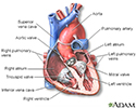
Heart - section through the middle - illustration
The interior of the heart is composed of valves, chambers, and associated vessels.
Heart - section through the middle
illustration
-
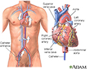
Cardiac catheterization - illustration
Cardiac catheterization is used to study the various functions of the heart. Using different techniques, the coronary arteries can be viewed by injecting dye or opened using balloon angioplasty. The oxygen concentration can be measured across the valves and walls (septa) of the heart and pressures within each chamber of the heart and across the valves can be measured. The technique can even be performed in small, newborn infants.
Cardiac catheterization
illustration
-
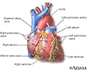
Heart - front view - illustration
The external structures of the heart include the ventricles, atria, arteries and veins. Arteries carry blood away from the heart while veins carry blood into the heart. The vessels colored blue indicate the transport of blood with relatively low content of oxygen and high content of carbon dioxide. The vessels colored red indicate the transport of blood with relatively high content of oxygen and low content of carbon dioxide.
Heart - front view
illustration
-
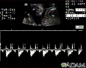
Ultrasound, normal fetus - heartbeat - illustration
This is a normal fetal ultrasound showing one pattern of the fetal heartbeat. Some ultrasound machines have the ability to focus on different areas of the heart and evaluate the heartbeat. This is useful in the early diagnosis of congenital heart abnormalities.
Ultrasound, normal fetus - heartbeat
illustration
-
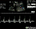
Ultrasound, ventricular septal defect - heartbeat - illustration
This is an ultrasound showing a ventricular septal defect pattern of the fetal heartbeat. Some ultrasound machines have the ability to focus on different areas of the heart and evaluate the heartbeat. This is useful in the early diagnosis of congenital heart abnormalities.
Ultrasound, ventricular septal defect - heartbeat
illustration
-
Patent ductus arteriosis (PDA) - series - Infant heart anatomy
Presentation
-
Infant open heart surgery - illustration
During open-heart surgery an incision is made through the breastbone (sternum) while the child is under general anesthesia.
Infant open heart surgery
illustration

 - series - Infant heart anatomy</strong><p>The heart pumps blood throughout the body. It is located in the thorax.</p>")
; Problems with heart rate or rhythm (arrhythmias); Excessive work load on heart that interferes with breathing, feeding, or sleeping.</p>")
 and between the lungs (mediastinum) while the child is deep asleep and pain-free (under general anesthesia). For some heart defect repairs, the incision is made on the side of the chest, between the ribs (thoracotomy) instead of through the breastbone. Heart lung bypass is used to support the child during the procedure. Tubes are used to re-route the blood through a special pump that adds oxygen to the blood and keeps it warm and moving through the rest of the body while the repair is being done.</p>")


-
Heart - section through the middle - illustration
The interior of the heart is composed of valves, chambers, and associated vessels.
Heart - section through the middle
illustration
-
Cardiac catheterization - illustration
Cardiac catheterization is used to study the various functions of the heart. Using different techniques, the coronary arteries can be viewed by injecting dye or opened using balloon angioplasty. The oxygen concentration can be measured across the valves and walls (septa) of the heart and pressures within each chamber of the heart and across the valves can be measured. The technique can even be performed in small, newborn infants.
Cardiac catheterization
illustration
-
Heart - front view - illustration
The external structures of the heart include the ventricles, atria, arteries and veins. Arteries carry blood away from the heart while veins carry blood into the heart. The vessels colored blue indicate the transport of blood with relatively low content of oxygen and high content of carbon dioxide. The vessels colored red indicate the transport of blood with relatively high content of oxygen and low content of carbon dioxide.
Heart - front view
illustration
-
Ultrasound, normal fetus - heartbeat - illustration
This is a normal fetal ultrasound showing one pattern of the fetal heartbeat. Some ultrasound machines have the ability to focus on different areas of the heart and evaluate the heartbeat. This is useful in the early diagnosis of congenital heart abnormalities.
Ultrasound, normal fetus - heartbeat
illustration
-
Ultrasound, ventricular septal defect - heartbeat - illustration
This is an ultrasound showing a ventricular septal defect pattern of the fetal heartbeat. Some ultrasound machines have the ability to focus on different areas of the heart and evaluate the heartbeat. This is useful in the early diagnosis of congenital heart abnormalities.
Ultrasound, ventricular septal defect - heartbeat
illustration
-
Patent ductus arteriosis (PDA) - series - Infant heart anatomy
Presentation
-
Infant open heart surgery - illustration
During open-heart surgery an incision is made through the breastbone (sternum) while the child is under general anesthesia.
Infant open heart surgery
illustration
Review Date: 1/23/2023
Reviewed By: Mary C. Mancini, MD, PhD, Cardiothoracic Surgeon, Shreveport, LA. Review provided by VeriMed Healthcare Network. Internal review and update on 02/26/24 by David C. Dugdale, MD, Medical Director, Brenda Conaway, Editorial Director, and the A.D.A.M. Editorial team.
 All rights reserved.