


Peristalsis
Intestinal motilityPeristalsis is a series of muscle contractions. These contractions occur in your digestive tract. Peristalsis is also seen in the tubes that connect the kidneys to the bladder.
Peristalsis is an automatic and important process. It moves:
- Food through the digestive system
- Urine from the kidneys into the bladder
- Bile from the gallbladder into the duodenum
Bile
Bile is a fluid that is made and released by the liver and stored in the gallbladder. Bile helps with digestion. It breaks down fats into fatty acid...
 ImageRead Article Now Book Mark Article
Duodenum
The duodenum is the first part of the small intestine. It is located between the stomach and the middle part of the small intestine, or jejunum. Aft...
 ImageRead Article Now Book Mark Article
Peristalsis is a normal function of the body. It can sometimes be felt in your belly (abdomen) as gas moves along.
References
Hall JE, Hall ME. General principles of gastrointestinal function - motility, nervous control, and blood circulation. In: Hall JE, Hall ME, eds. Guyton and Hall Textbook of Medical Physiology. 14th ed. Philadelphia, PA: Elsevier; 2021:chap 63.
Merriam-Webster's Medical Dictionary. Peristalsis. www.merriam-webster.com/medical. Accessed July 18, 2022.
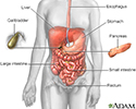
Digestive system - illustration
The esophagus, stomach, large and small intestine, aided by the liver, gallbladder and pancreas convert the nutritive components of food into energy and break down the non-nutritive components into waste to be excreted.
Digestive system
illustration
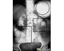
Ileus - X-ray of distended bowel and stomach - illustration
This abdominal X-ray shows a stomach filled with fluid and a swollen (distended) small bowel, caused by a blockage (pseudo-obstruction) in the intestines. A solution containing a dye (barium) that is visible on X-rays was swallowed by the patient (upper GI series).
Ileus - X-ray of distended bowel and stomach
illustration
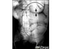
Ileus - X-ray of bowel distension - illustration
This abdominal X-ray shows thickening of the bowel wall and swelling (distention) caused by a blockage (pseudo-obstruction) in the intestines. A solution containing a dye (barium), which is visible on X-ray, was swallowed by the patient (the procedure is known as an upper GI series).
Ileus - X-ray of bowel distension
illustration
Peristalsis - illustration
A series of normal coordinated, rhythmic muscle contractions, that occurs automatically to move food through the digestive tract is called peristalsis.
Peristalsis
illustration

Digestive system - illustration
The esophagus, stomach, large and small intestine, aided by the liver, gallbladder and pancreas convert the nutritive components of food into energy and break down the non-nutritive components into waste to be excreted.
Digestive system
illustration
Ileus - X-ray of distended bowel and stomach - illustration
This abdominal X-ray shows a stomach filled with fluid and a swollen (distended) small bowel, caused by a blockage (pseudo-obstruction) in the intestines. A solution containing a dye (barium) that is visible on X-rays was swallowed by the patient (upper GI series).
Ileus - X-ray of distended bowel and stomach
illustration
Ileus - X-ray of bowel distension - illustration
This abdominal X-ray shows thickening of the bowel wall and swelling (distention) caused by a blockage (pseudo-obstruction) in the intestines. A solution containing a dye (barium), which is visible on X-ray, was swallowed by the patient (the procedure is known as an upper GI series).
Ileus - X-ray of bowel distension
illustration
Peristalsis - illustration
A series of normal coordinated, rhythmic muscle contractions, that occurs automatically to move food through the digestive tract is called peristalsis.
Peristalsis
illustration
Review Date: 7/25/2022
Reviewed By: Linda J. Vorvick, MD, Clinical Professor, Department of Family Medicine, UW Medicine, School of Medicine, University of Washington, Seattle, WA. Also reviewed by David C. Dugdale, MD, Medical Director, Brenda Conaway, Editorial Director, and the A.D.A.M. Editorial team.
 All rights reserved.