Aural polyps
Otic polypAn aural polyp is a growth in the outside (external) ear canal or middle ear. It may be attached to the eardrum (tympanic membrane), or it may grow from the middle ear space.
Causes
Aural polyps may be caused by:
- Cholesteatoma
Cholesteatoma
Cholesteatoma is a type of skin cyst that is located in the middle ear and mastoid bone in the skull.
 ImageRead Article Now Book Mark Article
 - Foreign object in the ear
- Inflammation of the ear canal or middle ear
- Tumor of the ear canal or middle ear
Symptoms
Bloody drainage from the ear is the most common symptom. Hearing loss can also occur.
Drainage from the ear
Ear discharge is drainage of blood, ear wax, pus, or fluid from the ear.

Exams and Tests
An aural polyp is diagnosed through an exam of the ear canal and middle ear using an otoscope or microscope.
Treatment
Treatment depends on the underlying cause. Your health care provider may first recommend:
- Avoiding water in the ear
- Steroid medicines
- Antibiotic ear drops
If a cholesteatoma is the underlying problem or the condition fails to clear, then surgery may be needed.
When to Contact a Medical Professional
Contact your health care provider if you have severe pain, bleeding from an ear or a sharp decrease in hearing.
References
Chi DH, Tobey A. Otolaryngology. In: Zitelli, BJ, McIntire SC, Nowalk AJ, Garrison J, eds. Zitelli and Davis' Atlas of Pediatric Physical Diagnosis. 7th ed. Philadelphia, PA: Elsevier; 2023:chap 24.
Chole RA, Sharon JD. Chronic otitis media, mastoiditis, and petrositis. In: Flint PW, Francis HW, Haughey BH, et al, eds. Cummings Otolaryngology: Head & Neck Surgery. 6th ed. Philadelphia, PA: Elsevier; 2021:chap 140.
McHugh JB. Ear. In: Goldblum JR, Lamps LW, McKenney JK, Myers JL, eds. Rosai and Ackerman's Surgical Pathology. 11th ed. Philadelphia, PA: Elsevier; 2018:chap 7.
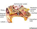
Ear anatomy - illustration
The ear consists of external, middle, and inner structures. The eardrum and the 3 tiny bones conduct sound from the eardrum to the cochlea.
Ear anatomy
illustration
Review Date: 5/2/2024
Reviewed By: Josef Shargorodsky, MD, MPH, Johns Hopkins University School of Medicine, Baltimore, MD. Also reviewed by David C. Dugdale, MD, Medical Director, Brenda Conaway, Editorial Director, and the A.D.A.M. Editorial team.


