Fusion of the ear bones
Fusion of the ear bones is an abnormal joining of the three bones of the middle ear. These are the incus, malleus, and stapes bones. Fusion or fixation of the bones leads to hearing loss because the bones are not moving and vibrating in reaction to sound waves.
Treatment options include hearing aids to amplify the sound and surgery to improve the middle ear conduction of sound.
Related topics include:
-
Chronic ear infection
Chronic ear infection
Chronic ear infection is fluid, swelling, or an infection behind the eardrum that does not go away or keeps coming back. It may cause long-term or p...
 ImageRead Article Now Book Mark Article
 -
Otosclerosis
Otosclerosis
Otosclerosis is an abnormal bone growth in the middle ear that causes hearing loss.
 ImageRead Article Now Book Mark Article
 - Middle ear malformations
References
House JW, Cunningham CD. Otosclerosis. In: Flint PW, Francis HW, Haughey BH, et al, eds. Cummings Otolaryngology: Head & Neck Surgery. 7th ed. Philadelphia, PA: Elsevier; 2021:chap 146.
O'Handley JG, Tobin EJ, Shah AR. Otorhinolaryngology. In: Rakel RE, Rakel DP, eds. Textbook of Family Medicine. 9th ed. Philadelphia, PA: Elsevier; 2016:chap 18.
Prueter JC, Teasley RA, Backous DD. Clinical assessment and surgical treatment of conductive hearing loss. In: Flint PW, Francis HW, Haughey BH, et al, eds. Cummings Otolaryngology: Head & Neck Surgery. 7th ed. Philadelphia, PA: Elsevier; 2021:chap 145.
Rivero A, Yoshikawa N. Otosclerosis. In: Myers EN, Snyderman CH, eds. Operative Otolaryngology Head and Neck Surgery. 3rd ed. Philadelphia, PA: Elsevier; 2018:chap 133.
-
Ear anatomy - illustration
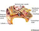
The ear consists of external, middle, and inner structures. The eardrum and the 3 tiny bones conduct sound from the eardrum to the cochlea.
Ear anatomy
illustration
-
Medical findings based on ear anatomy - illustration
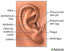
The external structures of the ear may aid in diagnosing some conditions by the presence or absence of normal landmarks and abnormal features including earlobe creases, preauricular pits, and preauricular tags.
Medical findings based on ear anatomy
illustration
-
Rheumatoid arthritis - InDepth
(In-Depth)
Review Date: 5/2/2024
Reviewed By: Josef Shargorodsky, MD, MPH, Johns Hopkins University School of Medicine, Baltimore, MD. Also reviewed by David C. Dugdale, MD, Medical Director, Brenda Conaway, Editorial Director, and the A.D.A.M. Editorial team.


