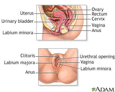
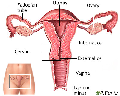
Retroversion of the uterus
Retroversion of the uterus occurs when a woman's uterus (womb) tilts backward rather than forward. It is commonly called a "tipped uterus."
Causes
Retroversion of the uterus is common. Approximately 1 in 5 women has this condition. The problem may also occur due to weakening of the pelvic ligaments at the time of menopause.
Menopause
Menopause is the time in a woman's life when her periods (menstruation) stop. Most often, it is a natural, normal body change that occurs between ag...

Scar tissue or adhesions in the pelvis can also hold the uterus in a retroverted position. Scarring may come from:
-
Endometriosis
Endometriosis
Endometriosis occurs when cells from the lining of your womb (uterus) grow in other areas of your body. This can cause pain, heavy vaginal bleeding,...
 ImageRead Article Now Book Mark Article
 -
Infection in uterus or tubes
Infection in uterus or tubes
Pelvic inflammatory disease (PID) is an infection of a woman's womb (uterus), ovaries, or fallopian tubes.
ImageRead Article Now Book Mark Article - Pelvic surgery
Symptoms
Retroversion of the uterus almost never causes any symptoms.
Rarely, it may cause pain or discomfort.
Exams and Tests
A pelvic exam will show the position of the uterus. However, a tipped uterus can sometimes be mistaken for a pelvic mass or a growing fibroid. A rectovaginal exam may be used to distinguish between a mass and a retroverted uterus.
Fibroid
Uterine fibroids are tumors that grow in a woman's womb (uterus). These growths are typically not cancerous (benign), and do not become cancerous....
An ultrasound exam can accurately determine the exact position of the uterus.
Ultrasound
Ultrasound uses high-frequency sound waves to make images of organs and structures inside the body.

Treatment
Treatment is not needed most of the time. Underlying disorders, such as endometriosis or adhesions, should be treated as needed.
Outlook (Prognosis)
In most cases, the condition does not cause problems.
Possible Complications
In most cases, a retroverted uterus is a normal finding. However, in some cases it may be caused by endometriosis, salpingitis, or pressure from a growing tumor.
When to Contact a Medical Professional
Call your health care provider if you have ongoing pelvic pain or discomfort.
Prevention
There is no way to prevent the problem. Early treatment of uterine infections or endometriosis may reduce the chances of a change in the position of the uterus.
Reviewed By
John D. Jacobson, MD, Department of Obstetrics and Gynecology, Loma Linda University School of Medicine, Loma Linda, CA. Also reviewed by David C. Dugdale, MD, Medical Director, Brenda Conaway, Editorial Director, and the A.D.A.M. Editorial team.
Advincula A, Truong M, Lobo RA. Endometriosis: etiology, pathology, diagnosis, management. In: Gershenson DM, Lentz GM, Valea FA, Lobo RA, eds. Comprehensive Gynecology. 8th ed. Philadelphia, PA: Elsevier; 2022:chap 19.
Ball JW, Dains JE, Flynn JA, Solomon BS, Stewart RW. Female genitalia. In: Ball JW, Dains JE, Flynn JA, Solomon BS, Stewart RW, eds. Seidel's Guide to Physical Examination. 10th ed. St Louis, MO: Elsevier; 2023:chap 19.
Hertzberg BS, Middleton WD. Pelvis and uterus. In: Hertzberg BS, Middleton WD, eds. Ultrasound: The Requisites. 3rd ed. Philadelphia, PA: Elsevier; 2016:chap 23.
Disclaimer
 All rights reserved.