


Leukemia
Leukemia is a type of blood cancer that begins in the bone marrow. Bone marrow is the soft tissue in the center of the bones, where blood cells are produced.
The term leukemia means white blood. White blood cells (leukocytes) are used by the body to fight infections and other foreign substances. Leukocytes are made in the bone marrow.
Leukemia leads to an uncontrolled increase in the number of white blood cells.
The cancerous cells prevent healthy red cells, platelets, and mature white cells (leukocytes) from being made. Life-threatening symptoms can then develop as normal blood cells decline.
The cancer cells can spread to the bloodstream and lymph nodes. They can also travel to the brain and spinal cord (the central nervous system) and other parts of the body.
Leukemia can affect children and adults.
Leukemias are divided into two major types:
- Acute (which progresses quickly)
- Chronic (which progresses more slowly)
The main types of leukemia are:
- Acute lymphocytic leukemia (ALL)
Acute lymphocytic leukemia
Acute lymphoblastic leukemia (ALL) is a fast-growing cancer of a type of white blood cells called lymphocytes. ALL occurs when the bone marrow produ...
 ImageRead Article Now Book Mark Article
 - Acute myelogenous leukemia (AML)
Acute myelogenous leukemia
Acute myeloid leukemia (AML) is cancer that starts inside bone marrow. This is the soft tissue in the center of bones that helps form all blood cell...
 ImageRead Article Now Book Mark Article
 - Chronic lymphocytic leukemia (CLL)
Chronic lymphocytic leukemia
Chronic lymphocytic leukemia (CLL) is cancer of a type of white blood cells called lymphocytes. These cells are found in the bone marrow and other p...
ImageRead Article Now Book Mark Article - Chronic myelogenous leukemia (CML)
Chronic myelogenous leukemia
Chronic myelogenous leukemia (CML) is cancer that starts inside bone marrow. This is the soft tissue in the center of bones that helps form all bloo...
ImageRead Article Now Book Mark Article
References
Appelbaum FR. Acute leukemias in adults. In: Niederhuber JE, Armitage JO, Kastan MB, Doroshow JH, Tepper JE, eds. Abeloff's Clinical Oncology. 6th ed. Philadelphia, PA: Elsevier; 2020:chap 95.
Hunger SP, Teachey DT, Grupp S, Aplenc R. Childhood leukemia. In: Niederhuber JE, Armitage JO, Kastan MB, Doroshow JH, Tepper JE, eds. Abeloff's Clinical Oncology. 6th ed. Philadelphia, PA: Elsevier; 2020:chap 93.
Bone marrow aspiration - illustration
A small amount of bone marrow is removed during a bone marrow aspiration. The procedure is uncomfortable, but can be tolerated by both children and adults. The marrow can be studied to determine the cause of anemia, the presence of leukemia or other malignancy, or the presence of some storage diseases, in which abnormal metabolic products are stored in certain bone marrow cells.
Bone marrow aspiration
illustration
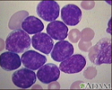
Acute lymphocytic leukemia - photomicrograph - illustration
This picture shows the darkly-stained lymph cells (lymphoblasts) seen in acute lymphocytic leukemia (ALL), the most common type of childhood leukemia.
Acute lymphocytic leukemia - photomicrograph
illustration
Auer rods - illustration
Note multiple Auer rods which are found only in acute myeloid leukemias, either myeloblastic or monoblastic. These rods consist of clumps of azurophilic granule material.
Auer rods
illustration
Chronic lymphocytic leukemia - microscopic view - illustration
This is a microscopic view of bone marrow from a person with chronic lymphocytic leukemia. It shows predominantly small, mature lymphocytes.
Chronic lymphocytic leukemia - microscopic view
illustration
Chronic myelocytic leukemia - microscopic view - illustration
This high-power microscopic view of a blood smear from a person with classical CML shows predominantly normal-appearing cells with intermediate maturity.
Chronic myelocytic leukemia - microscopic view
illustration
Chronic myelocytic leukemia - illustration
Oil immersion field demonstrating myeloid cells of all degrees of maturity.
Chronic myelocytic leukemia
illustration
Chronic myelocytic leukemia - illustration
Low power view showing marked hypercellularity with a broad-spectrum of myeloid and erythroid cell types and marked myeloid hyperplasia.
Chronic myelocytic leukemia
illustration


Bone marrow aspiration - illustration
A small amount of bone marrow is removed during a bone marrow aspiration. The procedure is uncomfortable, but can be tolerated by both children and adults. The marrow can be studied to determine the cause of anemia, the presence of leukemia or other malignancy, or the presence of some storage diseases, in which abnormal metabolic products are stored in certain bone marrow cells.
Bone marrow aspiration
illustration
Acute lymphocytic leukemia - photomicrograph - illustration
This picture shows the darkly-stained lymph cells (lymphoblasts) seen in acute lymphocytic leukemia (ALL), the most common type of childhood leukemia.
Acute lymphocytic leukemia - photomicrograph
illustration
Auer rods - illustration
Note multiple Auer rods which are found only in acute myeloid leukemias, either myeloblastic or monoblastic. These rods consist of clumps of azurophilic granule material.
Auer rods
illustration
Chronic lymphocytic leukemia - microscopic view - illustration
This is a microscopic view of bone marrow from a person with chronic lymphocytic leukemia. It shows predominantly small, mature lymphocytes.
Chronic lymphocytic leukemia - microscopic view
illustration
Chronic myelocytic leukemia - microscopic view - illustration
This high-power microscopic view of a blood smear from a person with classical CML shows predominantly normal-appearing cells with intermediate maturity.
Chronic myelocytic leukemia - microscopic view
illustration
Chronic myelocytic leukemia - illustration
Oil immersion field demonstrating myeloid cells of all degrees of maturity.
Chronic myelocytic leukemia
illustration
Chronic myelocytic leukemia - illustration
Low power view showing marked hypercellularity with a broad-spectrum of myeloid and erythroid cell types and marked myeloid hyperplasia.
Chronic myelocytic leukemia
illustration
Review Date: 1/25/2022
Reviewed By: Todd Gersten, MD, Hematology/Oncology, Florida Cancer Specialists & Research Institute, Wellington, FL. Review provided by VeriMed Healthcare Network. Also reviewed by David Zieve, MD, MHA, Medical Director, Brenda Conaway, Editorial Director, and the A.D.A.M. Editorial team.
 All rights reserved.