Sertoli-Leydig cell tumor
Sertoli-stromal cell tumor; Arrhenoblastoma; Androblastoma; Ovarian cancer - Sertoli-Leydig cell tumorSertoli-Leydig cell tumor (SLCT) is a rare cancer of the ovaries. The cancer cells produce and release a male sex hormone called testosterone.
Causes
The exact cause of this tumor is not known. Changes (mutations) in genes may play a role.
SLCT occur most often in young women 20 to 30 years old. But the tumor can occur at any age.
Symptoms
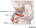
The Sertoli cells are normally located in the male reproductive glands (the testes). They feed sperm cells. The Leydig cells, also located in the testes, release a male sex hormone.
These cells are also found in a woman's ovaries, and in very rare cases lead to cancer. SLCT starts in the female ovaries, mostly in one ovary. The cancer cells release a male sex hormone. As a result, the woman may develop symptoms such as:
- A deep voice
- Enlarged clitoris
- Facial hair
Facial hair
Most of the time, women have fine hair above their lips and on their chin, chest, abdomen, or back. The growth of coarse dark hair in these areas (m...
Read Article Now Book Mark Article - Loss in breast size
- Stopping of menstrual periods
Pain in the lower belly (pelvic area) is another symptom. It occurs due to the tumor pressing on nearby structures.
Exams and Tests
The health care provider will perform a physical exam and a pelvic exam, and ask about the symptoms.
Tests will be ordered to check the levels of female and male hormones, including testosterone.
Testosterone
A testosterone test measures the amount of the male hormone, testosterone, in the blood. Both men and women produce this hormone. The test described...
Read Article Now Book Mark ArticleAn ultrasound or CT scan will likely be done to find out where the tumor is and its size and shape.
Ultrasound
A pelvic (transabdominal) ultrasound is an imaging test. It is used to examine organs in the pelvis.
Read Article Now Book Mark ArticleTreatment
Surgery is done to remove one or both ovaries.
If the tumor is advanced stage, chemotherapy or radiation therapy may be done after surgery.
Chemotherapy
The term chemotherapy is used to describe cancer-killing drugs. Chemotherapy may be used to:Cure the cancerShrink the cancerPrevent the cancer from ...

Radiation therapy
Radiation therapy uses high-powered radiation (such as x-rays or gamma rays), particles, or radioactive seeds to kill cancer cells.

Outlook (Prognosis)
Early treatment results in a good outcome. Feminine characteristics usually return after surgery. But male characteristics resolve more slowly.
For more advanced stage tumors, outlook is less positive.
References
Fletcher CDM. Tumors of the female genital tract. In: Fletcher CDM, ed. Diagnostic Histopathology of Tumors. 5th ed. Philadelphia, PA: Elsevier; 2021:chap 13.
Penick ER, Hamilton CA, Maxwell GL, Marcus CS. Germ cell, stromal, and other ovarian tumors. In: DiSaia PJ, Creasman WT, Mannel RS, McMeekin DS, Mutch DG, eds. Clinical Gynecologic Oncology. 9th ed. Philadelphia, PA: Elsevier; 2018:chap 12.
Smith RP. Sertoli-Leydig cell tumor (arrhenoblastoma). In: Smith RP, ed. Netter's Obstetrics & Gynecology. 3rd ed. Philadelphia, PA: Elsevier; 2018:chap 158.
Review Date: 4/29/2022
Reviewed By: Todd Gersten, MD, Hematology/Oncology, Florida Cancer Specialists & Research Institute, Wellington, FL. Review provided by VeriMed Healthcare Network. Also reviewed by David C. Dugdale, MD, Medical Director, Brenda Conaway, Editorial Director, and the A.D.A.M. Editorial team.


