

Corneal ulcers and infections
Bacterial keratitis; Fungal keratitis; Acanthamoeba keratitis; Herpes simplex keratitisThe cornea is the clear tissue at the front of the eye. A corneal ulcer is an open sore in the outer layer of the cornea. It is often caused by infection. At first, a corneal ulcer may seem like conjunctivitis, or pink eye.
Causes
Corneal ulcers are most commonly caused by an infection with bacteria, viruses, fungi, or a parasite.
- Acanthamoeba keratitis occurs in contact lens users. It is more likely to happen in people who make their own homemade cleaning solutions.
- Fungal keratitis can occur after a corneal injury involving plant material. It may also occur in people with a suppressed immune system.
- Herpes simplex virus keratitis is a serious viral infection. It may cause repeated attacks that are triggered by stress, exposure to sunlight, or any condition that lowers the immune response.
Corneal ulcers or infections may also be caused by:
- Eyelids that do not close all the way, such as with Bell palsy
Bell palsy
Bell palsy is a disorder of the nerve that controls movement of the muscles in the face. This nerve is called the facial or seventh cranial nerve. D...
 ImageRead Article Now Book Mark Article
 - Foreign bodies in the eye
- Scratches (abrasions) on the eye surface
- Severely dry eyes
- Severe allergic eye disease
- Various inflammatory disorders
Wearing contact lenses, especially soft contacts that are left in overnight, may cause a corneal ulcer.
Symptoms
Symptoms of infections or ulcers of the cornea include:
- Blurry or hazy vision
- Eye that appears red or bloodshot
-
Itching and discharge
Itching and discharge
Eye burning with discharge is burning, itching, or drainage from the eye of any substance other than tears.
 ImageRead Article Now Book Mark Article
 -
Sensitivity to light (photophobia)
Sensitivity to light
Photophobia is eye discomfort in bright light.
ImageRead Article Now Book Mark Article - Very painful and watery eyes
Watery eyes
Watery eyes means you have too many tears in and draining from the eyes. Tears help keep the surface of the eye moist. They wash away particles and...
ImageRead Article Now Book Mark Article - White patch on the cornea
Exams and Tests
Your health care provider or eye doctor may do the following tests:
- Exam of scrapings from the ulcer
- Fluorescein stain of the cornea
- Keratometry (measuring the curve of the cornea)
- Pupillary reflex response
-
Refraction test
Refraction test
A refraction is an eye exam that measures a person's prescription for eyeglasses or contact lenses.
 ImageRead Article Now Book Mark Article
 -
Slit-lamp examination
Slit-lamp examination
The slit-lamp examination looks at structures that are at the front of the eye.
 ImageRead Article Now Book Mark Article
 - Tests for dry eye
-
Visual acuity
Visual acuity
The visual acuity test is used to determine the smallest letters you can read on a standardized chart (Snellen chart) or a card held 20 feet (6 meter...
ImageRead Article Now Book Mark Article
Blood tests to check for inflammatory disorders may also be needed.
Using newer information systems to evaluate photos of corneal ulcers may allow earlier diagnosis and treatment.
Treatment
Treatment for corneal ulcers and infections depends on the cause. Treatment should be started as soon as possible to prevent scarring of the cornea.
If the exact cause is not known, you may be given antibiotic drops that work against many kinds of bacteria.
Once the exact cause is known, you may be given drops that treat bacteria, herpes simplex virus, other viruses, or a fungus. Severe ulcers sometimes require a corneal transplant.
Corticosteroid eye drops may be used to reduce swelling and inflammation in certain conditions.
Your provider may also recommend that you:
- Avoid eye makeup.
- Do not wear contact lenses at all, especially while asleep.
- Take pain medicines.
- Wear protective glasses.
Outlook (Prognosis)
Many people recover completely and have only a minor change in vision. However, a corneal ulcer or infection can cause long-term damage and affect vision.
Possible Complications
Untreated corneal ulcers and infections may lead to:
- Loss of the eye (rare)
- Severe vision loss
- Scars on the cornea
When to Contact a Medical Professional
Contact your provider if:
- You have symptoms of corneal ulcers or an infection.
- You have been diagnosed with this condition and your symptoms become worse after treatment.
- Your vision is affected.
- You develop eye pain that is severe or becoming worse.
- Your eyelids or the skin around your eyes becomes swollen or red.
- You have a headache in addition to your other symptoms.
Prevention
Things you can do to prevent the condition include:
- Wash your hands well when handling your contact lenses.
- Avoid wearing contact lenses overnight.
- Get prompt treatment for an eye infection to prevent ulcers from forming.
References
Azar DT, Hallak J, Barnes SD, Giri P, Pavan-Langston D. Microbial keratitis. In: Bennett JE, Dolin R, Blaser MJ, eds. Mandell, Douglas, and Bennett's Principles and Practice of Infectious Diseases. 9th ed. Philadelphia, PA: Elsevier; 2020:chap 113.
Cioffi GA, Liebmann JM. Diseases of the visual system. In: Goldman L, Cooney KA, eds. Goldman-Cecil Medicine. 27th ed. Philadelphia, PA: Elsevier; 2024:chap 391.
Efron N. Corneal staining. In: Efron N, ed. Contact Lens Complications. 4th ed. Philadelphia, PA: Elsevier; 2019:chap 18.
Keenan JD, McLeod SD. Bacterial keratitis. In: Yanoff M, Duker JS, eds. Ophthalmology. 6th ed. Philadelphia, PA: Elsevier; 2023:chap 4.12.
Tuli SS, Steigleman WA. Herpes simplex keratitis. In: Yanoff M, Duker JS, eds. Ophthalmology. 6th ed. Philadelphia, PA: Elsevier; 2023:chap 4.15.
-
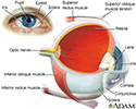
Eye - illustration
The eye is the organ of sight, a nearly spherical hollow globe filled with fluids (humors). The outer layer (sclera, or white of the eye, and cornea) is fibrous and protective. The middle layer (choroid, ciliary body and the iris) is vascular. The innermost layer (retina) is sensory nerve tissue that is light sensitive. The fluids in the eye are divided by the lens into the vitreous humor (behind the lens) and the aqueous humor (in front of the lens). The lens itself is flexible and suspended by ligaments which allow it to change shape to focus light on the retina, which is composed of sensory neurons.
Eye
illustration
-
Eye - illustration
The eye is the organ of sight, a nearly spherical hollow globe filled with fluids (humors). The outer layer (sclera, or white of the eye, and cornea) is fibrous and protective. The middle layer (choroid, ciliary body and the iris) is vascular. The innermost layer (retina) is sensory nerve tissue that is light sensitive. The fluids in the eye are divided by the lens into the vitreous humor (behind the lens) and the aqueous humor (in front of the lens). The lens itself is flexible and suspended by ligaments which allow it to change shape to focus light on the retina, which is composed of sensory neurons.
Eye
illustration
Review Date: 7/9/2024
Reviewed By: Audrey Tai, DO, MS, Athena Eye Care, Mission Viejo, CA. Also reviewed by David C. Dugdale, MD, Medical Director, Brenda Conaway, Editorial Director, and the A.D.A.M. Editorial team.
 All rights reserved.