

Hyphema
Hyphema is blood in the front area (anterior chamber) of the eye. The blood collects behind the cornea and in front of the iris.
Causes
Hyphema is most often caused by trauma to the eye. Other causes of bleeding in the front chamber of the eye include:
- Blood vessel abnormality
- Cancer of the eye
- Severe inflammation of the iris
Iris
The iris is the colored part of the eye. It is located between the cornea and lens. The round, central opening of the iris is called the pupil. Ve...
 ImageRead Article Now Book Mark Article
 - Advanced diabetes
Diabetes
Diabetes is a long-term (chronic) disease in which the body cannot regulate the amount of sugar in the blood.
 ImageRead Article Now Book Mark Article
 - Blood disorders such as sickle cell anemia
Sickle cell anemia
Sickle cell disease is a disorder passed down through families. The red blood cells that are normally shaped like a disk take on a sickle or crescen...
 ImageRead Article Now Book Mark Article
Symptoms
Symptoms include:
- Bleeding in the anterior chamber of the eye
-
Eye pain
Eye pain
Pain in the eye may be described as a burning, throbbing, aching, or stabbing sensation in or around the eye. It may also feel like you have a forei...
Read Article Now Book Mark Article - Light sensitivity
-
Vision abnormalities
Vision abnormalities
There are many types of eye problems and vision disturbances, such as: Halos Blurred vision (the loss of sharpness of vision and the inability to see...
 ImageRead Article Now Book Mark Article
You may not be able to see a small hyphema when looking at your eye in the mirror. With a total hyphema, the collection of blood will block the view of the iris and pupil.
Exams and Tests
You may need the following tests and exams:
- Eye exam
- Intraocular pressure measurement (tonometry)
Tonometry
Tonometry is a test to measure the pressure inside your eyes. The test is used to screen for glaucoma. It is also used to measure how well glaucoma...
 ImageRead Article Now Book Mark Article
 -
Ultrasound testing
Ultrasound
Ultrasound uses high-frequency sound waves to make images of organs and structures inside the body.
 ImageRead Article Now Book Mark Article
Treatment
Treatment may not be needed in mild cases. The blood is absorbed in a few days.
The outcome of the condition will likely be much worse if bleeding comes back or worsens, especially within 3 to 5 days. The health care provider may recommend the following to cut down the chance that there will be more bleeding:
- Bed rest
- Eye patching
- Sedating medicines
You may need to use eye drops to decrease the inflammation or lower the pressure in your eye.
The eye doctor may need to remove the blood surgically, especially if pressure in the eye is very high or the blood is slow to absorb again. You may need to stay in a hospital.
Outlook (Prognosis)
The outcome depends upon the amount of injury to the eye. People with sickle cell disease are more likely to have eye complications and must be watched closely. People with diabetes will probably need laser treatment for the problem.
Sickle cell disease
Sickle cell disease is a disorder passed down through families. The red blood cells that are normally shaped like a disk take on a sickle or crescen...
Severe vision loss can occur.
Possible Complications
Complications may include:
-
Acute glaucoma
Acute glaucoma
Glaucoma is a group of eye conditions that can damage the optic nerve. This nerve sends the images you see to your brain. Most often, optic nerve da...
ImageRead Article Now Book Mark Article - Impaired vision
- Recurring bleeding
When to Contact a Medical Professional
Call your provider if you notice blood in the front of the eye or if you have an eye injury. You will need to be examined and treated by an eye doctor right away, especially if you have decreased vision.
Prevention
Many eye injuries can be prevented by wearing safety goggles or other protective eye wear. Always wear eye protection while playing sports, such as racquetball, or contact sports, such as basketball.
References
Lin TKY, Tingey DP, Shingleton BJ. Glaucoma associated with ocular trauma. In: Yanoff M, Duker JS, eds. Ophthalmology. 6th ed. Philadelphia, PA: Elsevier; 2022:chap 10.16.
Olitsky SE, Marsh JD. Injuries to the eye. In: Kliegman RM, St. Geme JW, Blum NJ, Shah SS, Tasker RC, Wilson KM, eds. Nelson Textbook of Pediatrics. 21st ed. Philadelphia, PA: Elsevier; 2020:chap 653.
Patel S, Kim SJ, Sternberg P. Surgery for ocular trauma: principles and techniques of treatment. In: Sadda SVR, Sarraf D, Freund B et al, eds. Ryan's Retina. 7th ed. Philadelphia, PA: Elsevier; 2023:chap 113.
-
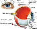
Eye - illustration
The eye is the organ of sight, a nearly spherical hollow globe filled with fluids (humors). The outer layer (sclera, or white of the eye, and cornea) is fibrous and protective. The middle layer (choroid, ciliary body and the iris) is vascular. The innermost layer (retina) is sensory nerve tissue that is light sensitive. The fluids in the eye are divided by the lens into the vitreous humor (behind the lens) and the aqueous humor (in front of the lens). The lens itself is flexible and suspended by ligaments which allow it to change shape to focus light on the retina, which is composed of sensory neurons.
Eye
illustration
-
Eye - illustration
The eye is the organ of sight, a nearly spherical hollow globe filled with fluids (humors). The outer layer (sclera, or white of the eye, and cornea) is fibrous and protective. The middle layer (choroid, ciliary body and the iris) is vascular. The innermost layer (retina) is sensory nerve tissue that is light sensitive. The fluids in the eye are divided by the lens into the vitreous humor (behind the lens) and the aqueous humor (in front of the lens). The lens itself is flexible and suspended by ligaments which allow it to change shape to focus light on the retina, which is composed of sensory neurons.
Eye
illustration
Review Date: 11/10/2022
Reviewed By: Franklin W. Lusby, MD, Ophthalmologist, Lusby Vision Institute, La Jolla, CA. Also reviewed by David C. Dugdale, MD, Medical Director, Brenda Conaway, Editorial Director, and the A.D.A.M. Editorial team.
 All rights reserved.