Eyelid bump
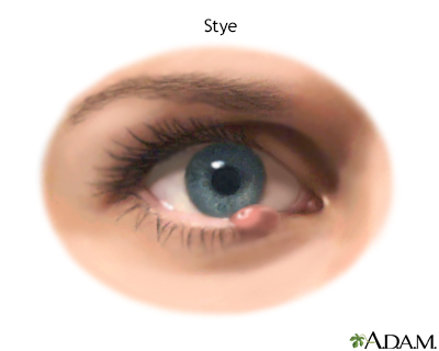
Bump on the eyelid; Stye; HordeolumMost bumps on the eyelid are styes. A stye is an inflamed oil gland on the edge of your eyelid, where the eyelash meets the lid. It appears as a red, swollen bump that looks like a pimple. It is often tender to the touch.
Causes
A stye is caused by a blockage of one of the oil glands in the eyelids. This allows bacteria to grow inside the blocked gland. Styes are a lot like common acne pimples that occur elsewhere on the skin. You may have more than one stye at the same time.
Styes most often develop over a few days. They may drain and heal on their own. A stye can become a chalazion, which occurs when an inflamed oil gland becomes fully blocked. If a chalazion gets large enough, it can cause trouble with your vision.
Chalazion
A chalazion is a small bump in the eyelid caused by a blockage of a tiny oil gland.

Styes can be made worse by the presence of Demodex, a mite commonly found on human skin. Demodex has been identified as a cause of blepharitis. If you have blepharitis, you are more likely to get styes.
Blepharitis
Blepharitis is manifest by inflamed, irritated, itchy, and reddened eyelids. It most often occurs where the eyelashes grow. Dandruff-like debris bu...

Other possible common eyelid bumps include:
- Xanthelasma: Raised yellow patches on your eyelids that can happen with age. These are harmless, although they are sometimes a sign of high cholesterol.
Xanthelasma
Xanthoma is a skin condition in which certain fats build up under the surface of the skin.
 ImageRead Article Now Book Mark Article
 - Papillomas: Pink or skin-colored bumps. They are harmless, but can slowly grow, affect your vision, or bother you for cosmetic reasons. If so, they can be surgically removed.
- Cysts: Small fluid-filled sacs that can affect your vision.
Symptoms
In addition to the red, swollen bump, other possible symptoms of a stye include:
- A gritty, scratchy sensation, as if there is a foreign body in your eye
- Sensitivity to light
Sensitivity
There are many types of eye problems and vision disturbances, such as: Halos Blurred vision (the loss of sharpness of vision and the inability to see...
 ImageRead Article Now Book Mark Article
 - Tearing of your eye
Tearing of your eye
Watery eyes means you have too many tears in and draining from the eyes. Tears help keep the surface of the eye moist. They wash away particles and...
 ImageRead Article Now Book Mark Article
 - Tenderness of the eyelid
Exams and Tests
In most cases, your health care provider can diagnose a stye just by looking at it. Tests are rarely needed.
Treatment
To treat eyelid bumps at home:
- Apply a warm, wet cloth to the area for 10 minutes. Do this 4 times a day.
- Do NOT attempt to squeeze a stye or any other type of eyelid bump. Let it drain on its own.
- Do NOT use contact lenses or wear eye makeup until the area has healed.
For a stye, your provider may:
- Prescribe antibiotic ointment
- Make an opening in the stye to drain it (Do NOT try this at home)
Outlook (Prognosis)
Styes often get better on their own. However, they may return. If bleparitis is present, treating it may help prevent a recurrence of the stye.
The outcome is almost always excellent with simple treatment.
Sometimes, the infection may spread to the rest of the eyelid. This is called eyelid cellulitis and may require oral antibiotics. This can look like orbital cellulitis, which can be serious problem, especially in children.
Orbital cellulitis
Orbital cellulitis is an infection of the fat and muscles around the eye. It affects the eyelids, eyebrows, and cheeks. It may begin suddenly or be...

When to Contact a Medical Professional
Contact your provider if:
- You have problems with your vision.
- The eyelid bump worsens or does not improve within a week or two of self-care.
- The eyelid bump or bumps become very large or painful.
- You have a blister on your eyelid.
- You have crusting or scaling of your eyelids.
- Your whole eyelid is red, or the eye itself is red.
- You are very sensitive to light or have excessive tears.
- Another stye comes back soon after successful treatment of a stye.
- Your eyelid bump bleeds.
Prevention
Always wash your hands very well before touching the skin around your eye. If you are prone to getting styes or have blepharitis, it may help to carefully clean off excess oils from the edges of your lids. To do this, use a solution of warm water and no-tears baby shampoo.
Fish oil taken by mouth may help prevent plugging of the oil glands. Treating for Demodex may also be helpful. An eye drop for this condition is also available. Your provider may also suggest using tea tree oil on the lids for Demodex.
References
Cioffi GA, Liebmann JM. Diseases of the visual system. In: Goldman L, Cooney KA, eds. Goldman-Cecil Medicine. 27th ed. Philadelphia, PA: Elsevier; 2024:chap 391.
Dupre AA, Voita LR. Red and painful eye. In: Walls RM, ed. Rosen's Emergency Medicine: Concepts and Clinical Practice. 10th ed. Philadelphia, PA: Elsevier; 2023:chap 18.
Neff AG, Chahal HS, Carter KD. Benign eyelid lesions. In: Yanoff M, Duker JS, eds. Ophthalmology. 6th ed. Philadelphia, PA: Elsevier; 2023:chap 12.7.
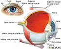
Eye - illustration
The eye is the organ of sight, a nearly spherical hollow globe filled with fluids (humors). The outer layer (sclera, or white of the eye, and cornea) is fibrous and protective. The middle layer (choroid, ciliary body and the iris) is vascular. The innermost layer (retina) is sensory nerve tissue that is light sensitive. The fluids in the eye are divided by the lens into the vitreous humor (behind the lens) and the aqueous humor (in front of the lens). The lens itself is flexible and suspended by ligaments which allow it to change shape to focus light on the retina, which is composed of sensory neurons.
Eye
illustration
Stye - illustration
A stye is a relatively common infection. Styes are often caused by staphylococcus and occur in the glands that open onto the lid margin. They are red, swollen, and painful.
Stye
illustration
Review Date: 7/9/2024
Reviewed By: Audrey Tai, DO, MS, Athena Eye Care, Mission Viejo, CA. Also reviewed by David C. Dugdale, MD, Medical Director, Brenda Conaway, Editorial Director, and the A.D.A.M. Editorial team.


