
Amaurosis fugax
Transient monocular blindness; Transient monocular visual loss; TMVL; Transient monocular visual loss; Transient binocular visual loss; TBVL; Temporary visual loss - amaurosis fugaxAmaurosis fugax is a temporary loss of vision in one or both eyes due to a lack of blood flow to the retina. The retina is the light-sensitive layer of tissue at the back of the eyeball.
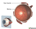
Retina
The retina is the light-sensitive layer of tissue at the back of the eyeball. Images that come through the eye's lens are focused on the retina. Th...

Causes
Amaurosis fugax is not itself a disease. Instead, it is a sign of other disorders. Amaurosis fugax can occur from different causes. One cause is when a blood clot or a piece of plaque blocks an artery in the eye. The blood clot or plaque usually travels from a larger artery, such as the carotid artery in the neck or an artery in the heart, to an artery in the eye.
Plaque is a hard substance that forms when fat, cholesterol, and other substances build up in the walls of arteries. Risk factors include:
- Heart disease, especially irregular heartbeat
- Alcohol abuse
Alcohol abuse
Alcohol use disorder is when your drinking causes serious problems in your life, yet you keep drinking. You may also need more and more alcohol to f...
 ImageRead Article Now Book Mark Article
 - Cocaine use
- Diabetes
Diabetes
Diabetes is a long-term (chronic) disease in which the body cannot regulate the amount of sugar in the blood.
 ImageRead Article Now Book Mark Article
 - Family history of stroke
Stroke
A stroke occurs when blood flow to a part of the brain stops. A stroke is sometimes called a "brain attack. " If blood flow is cut off for longer th...
 ImageRead Article Now Book Mark Article
 - High blood pressure
- High cholesterol
- Increasing age
- Smoking (people who smoke one pack a day double their risk for a stroke)
Amaurosis fugax can also occur because of other disorders such as:
- Other eye problems, such as inflammation of the optic nerve (optic neuritis)
Optic neuritis
The optic nerve carries images of what the eye sees to the brain. When this nerve become swollen or inflamed, it is called optic neuritis. It may c...
 ImageRead Article Now Book Mark Article
 - Blood vessel disease called polyarteritis nodosa
Polyarteritis nodosa
Polyarteritis nodosa is a serious inflammatory blood vessel disease. The small and medium-sized arteries become swollen and damaged.
 ImageRead Article Now Book Mark Article
 - Migraine headaches
Migraine headaches
A migraine is a type of headache. It may occur with symptoms such as nausea, vomiting, or sensitivity to light and sound. In most people, a throbbi...
 ImageRead Article Now Book Mark Article
 - Brain tumor
- Head injury
- Multiple sclerosis (MS), inflammation of the nerves due to the body's immune cells attacking the nervous system
Multiple sclerosis
Multiple sclerosis (MS) is an autoimmune disease that affects the brain and spinal cord (central nervous system).
 ImageRead Article Now Book Mark Article
 - Systemic lupus erythematosus, an autoimmune disease in which the body's immune cells attack healthy tissue throughout the body
Systemic lupus erythematosus
Systemic lupus erythematosus (SLE) is an autoimmune disease. In this disease, the immune system of the body mistakenly attacks healthy tissue. It c...
 ImageRead Article Now Book Mark Article
Symptoms
Symptoms include the sudden loss of vision in one or both eyes. This usually lasts for a few seconds to several minutes. Afterward, vision returns to normal. Some people describe the loss of vision as a gray or black shade coming down over the eye.
Exams and Tests
Your health care provider will perform a complete eye and nervous system exam. In some cases, an eye exam will reveal a bright spot where the clot is blocking a retinal artery.
Tests that may be done include:
- Ultrasound or magnetic resonance angiography scan of the carotid artery to check for blood clots or plaque
Ultrasound
Carotid duplex is an ultrasound test that shows how well blood is flowing through the carotid arteries. The carotid arteries are located in the neck...
 ImageRead Article Now Book Mark Article
Magnetic resonance angiography scan
Magnetic resonance angiography (MRA) is an MRI exam of the blood vessels. Unlike traditional angiography that involves placing a tube (catheter) int...
 ImageRead Article Now Book Mark Article
 - Blood tests to check cholesterol and blood sugar levels
- Tests of the heart, such as an electrocardiogram (ECG), or Holter monitor or loop recorder to check its electrical activity
Electrocardiogram
An electrocardiogram (ECG) is a test that records the electrical activity of the heart.
 ImageRead Article Now Book Mark Article
 - Echocardiogram to check the structure of the heart and to look for blood clots in the heart
Echocardiogram
An echocardiogram is a test that uses sound waves to create pictures of the heart. The picture and information it produces is more detailed than a s...
 ImageRead Article Now Book Mark Article
Treatment
Treatment of amaurosis fugax depends on its cause. When amaurosis fugax is due to a blood clot or plaque, the concern is to prevent a stroke. The following can help prevent a stroke:
- Avoid fatty foods and follow a healthy, low-fat diet. Do not drink more than 1 to 2 alcoholic drinks a day.
- Exercise regularly: 30 minutes a day if you are not overweight; 60 to 90 minutes a day if you are overweight.
- Quit smoking.
Quit smoking
There are many ways to quit smoking. There are also resources to help you. Family members, friends, and co-workers may be supportive. But to be su...
 ImageRead Article Now Book Mark Article
 - Most people should aim for a blood pressure below 120 to 130/80 mm Hg. If you have diabetes or have had a stroke, your provider may tell you to aim for a lower blood pressure.
- If you have diabetes, heart disease, or hardening of the arteries, your LDL (bad) cholesterol should be lower than 70 mg/dL. Some experts recommend an even lower target, below 55 mg/dL.
- Follow your provider's treatment plans if you have high blood pressure, diabetes, high cholesterol, or heart disease.
Your provider may also recommend:
- No treatment. You may only need regular visits to check the health of your heart and carotid arteries.
- Aspirin, warfarin (Coumadin), or other blood-thinning medicines to lower your risk for stroke.
If a large part of the carotid artery appears blocked, carotid endarterectomy surgery is done to remove the blockage. The decision to do surgery is also based on your overall health.
Carotid endarterectomy
Carotid artery surgery is a procedure to treat carotid artery disease. The carotid artery brings needed blood to your brain and face. You have one o...

Outlook (Prognosis)
Amaurosis fugax increases your risk for stroke.
When to Contact a Medical Professional
Contact your provider if any vision loss occurs. If symptoms last longer than a few minutes or if there are other symptoms with the vision loss, seek medical attention right away.
References
Biller J, Schneck MJ, Ruland S. Ischemic cerebrovascular disease. In: Jankovic J, Mazziotta JC, Pomeroy SL, Newman NJ, eds. Bradley and Daroff's Neurology in Clinical Practice. 8th ed. Philadelphia, PA: Elsevier; 2022:chap 65.
Brown GC, Sharma S, Brown MM. Ocular ischemic syndrome. In: Sadda SVR, Sarraf D, Freund KB, et al, eds. Ryan's Retina. 7th ed. Philadelphia, PA: Elsevier; 2023:chap 61.
Lloyd-Jones DM, Morris PB, Ballantyne CM, et al. 2022 ACC expert consensus decision pathway on the role of nonstatin therapies for LDL-Cholesterol lowering in the management of atherosclerotic cardiovascular disease risk: A Report of the American College of Cardiology Solution Set Oversight Committee. J Am Coll Cardiol. 2022;80(14):1366-1418. PMID: 36031461 pubmed.ncbi.nlm.nih.gov/36031461/.
Retina - illustration
The retina is the internal layer of the eye that receives and transmits focused images. The retina is normally red due to its rich blood supply.
Retina
illustration
Review Date: 6/13/2024
Reviewed By: Joseph V. Campellone, MD, Department of Neurology, Cooper Medical School at Rowan University, Camden, NJ. Review provided by VeriMed Healthcare Network. Also reviewed by David C. Dugdale, MD, Medical Director, Brenda Conaway, Editorial Director, and the A.D.A.M. Editorial team.
 All rights reserved.