Horner syndrome
Oculosympathetic paresis; Horner's syndromeHorner syndrome is a rare condition that affects the nerves to the eye and face.
Causes
Horner syndrome can be caused by any interruption in a set of nerve fibers that start in the part of the brain called the hypothalamus and travel to the face and eyes. These nerve fibers are involved with sweating, the pupils in your eyes, and the upper and lower eyelid muscles.
Damage of the nerve fibers can result from:
- Injury to the carotid artery, one of the main arteries to the brain
- Injury to nerves at the base of the neck called the brachial plexus
-
Migraine or cluster headaches
Migraine
A migraine is a type of headache. It may occur with symptoms such as nausea, vomiting, or sensitivity to light and sound. In most people, a throbbi...
 ImageRead Article Now Book Mark Article
Cluster
A cluster headache is an uncommon type of headache. It is one-sided head pain that may involve tearing of the eyes, a droopy eyelid, and a stuffy no...
 ImageRead Article Now Book Mark Article
 -
Stroke, tumor, or other damage to a part of the brain called the brainstem
Stroke
A stroke occurs when blood flow to a part of the brain stops. A stroke is sometimes called a "brain attack. " If blood flow is cut off for longer th...
 ImageRead Article Now Book Mark Article
 - Tumor or infection in the top of the lung, between the lungs, and neck
- Injections or surgery done to interrupt the nerve fibers and relieve pain (sympathectomy)
-
Spinal cord injury
Spinal cord injury
Spinal cord trauma is damage to the spinal cord. It may result from direct injury to the cord itself or indirectly from disease of the nearby bones,...
 ImageRead Article Now Book Mark Article
In rare cases, Horner syndrome is present at birth. The condition may occur with a lack of color (pigmentation) of the iris (colored part of the eye).
Symptoms
Symptoms of Horner syndrome may include:
- Decreased sweating on the affected side of the face
-
Drooping eyelid (ptosis)
Drooping eyelid
Ptosis (eyelid drooping) in infants and children is when the upper eyelid is lower than it should be. This may occur in one or both eyes. Eyelid dr...
 ImageRead Article Now Book Mark Article
Ptosis
Eyelid drooping is excess sagging of the upper eyelid. The edge of the upper eyelid may be lower than it should be (ptosis) or there may be excess b...
 ImageRead Article Now Book Mark Article
 - Sinking of the eyeball into the face
- Different sizes of pupils of the eyes (anisocoria) with the affected side pupil being smaller
Anisocoria
Anisocoria is unequal pupil size. The pupil is the black part in the center of the eye. It gets larger in dim light and smaller in bright light....
 ImageRead Article Now Book Mark Article
There may also be other symptoms, depending on the location of the affected nerve fiber. These may include:
-
Vertigo (sensation that surroundings are spinning) with nausea and vomiting
Vertigo
Vertigo is a sensation of motion or spinning that is often described as dizziness. Vertigo is not the same as being lightheaded. People with vertigo...
 ImageRead Article Now Book Mark Article
 - Double vision
- Lack of muscle control and coordination
- Arm pain, weakness and numbness
- One sided neck and ear pain
-
Hoarseness
Hoarseness
Hoarseness refers to difficulty making sounds when trying to speak. Vocal sounds may be weak, breathy, scratchy, or husky, and the pitch or quality ...
 ImageRead Article Now Book Mark Article
 - Hearing loss
- Bladder and bowel difficulty
- Overreaction of the involuntary (autonomic) nervous system to stimulation (hyperreflexia)
Hyperreflexia
Autonomic dysreflexia is an abnormal, overreaction of the involuntary (autonomic) nervous system to stimulation. This reaction may include: Change i...
 ImageRead Article Now Book Mark Article
Exams and Tests
The health care provider will perform a physical exam and ask about the symptoms.
An eye exam may show:
- Changes in how the pupil opens or closes
- Constricted pupil
- Eyelid drooping
- Red eye
Depending on the suspected cause, tests may be done, such as:
- Blood tests
- Blood vessel tests of the head (angiogram)
Angiogram
An arteriogram is an imaging test that uses x-rays and a special dye to see inside the arteries. It can be used to view arteries in the heart, brain...
 ImageRead Article Now Book Mark Article
 -
Chest x-ray or chest CT scan
Chest x-ray
A chest x-ray is an x-ray of the chest, lungs, heart, large arteries, ribs, and diaphragm.
 ImageRead Article Now Book Mark Article
Chest CT scan
A chest CT (computed tomography) scan is an imaging method that uses x-rays to create cross-sectional pictures of the chest and upper abdomen....
 ImageRead Article Now Book Mark Article
 -
MRI or CT scan of the brain or cervical spine
MRI
A head MRI (magnetic resonance imaging) is an imaging test that uses powerful magnets and radio waves to create pictures of the brain and surrounding...
 ImageRead Article Now Book Mark Article
CT scan of the brain
A head computed tomography (CT) scan uses many x-rays to create pictures of the head, including the skull, brain, eye sockets, and sinuses.
 ImageRead Article Now Book Mark Article
 -
Spinal tap (lumbar puncture)
Spinal tap
Cerebrospinal fluid (CSF) collection is a test to look at the fluid that surrounds the brain and spinal cord. CSF acts as a cushion, protecting the b...
 ImageRead Article Now Book Mark Article
 - Electromyography and nerve conduction studies if nerve root or brachial plexus injury suspected
You may need to be referred to a doctor who specializes in vision problems related to the nervous system (neuro-ophthalmologist).
Treatment
Treatment depends on the underlying cause of the condition. There is no treatment for Horner syndrome itself. Ptosis is very mild and in rare cases affects vision in Horner syndrome. This can be corrected by cosmetic surgery or treated with eyedrops. The provider can tell you more.
Outlook (Prognosis)
The outcome depends on whether treatment of the cause is successful.
Possible Complications
There are no direct complications of Horner syndrome itself. But, there may be complications from the disease that caused Horner syndrome or from its treatment.
When to Contact a Medical Professional
Call your provider if you have symptoms of Horner syndrome.
References
Balcer LJ. Pupillary disorders. In: Liu GT, Volpe NJ, Galetta SL, eds. Liu, Volpe, and Galetta's Neuro-Ophthalmology. 3rd ed. Philadelphia, PA: Elsevier; 2019:chap 13.
Norse AB. Diplopia. In: Walls RM, ed. Rosen's Emergency Medicine: Concepts and Clinical Practice. 10th ed. Philadelphia, PA: Elsevier; 2023:chap 17.
Thurtell MJ, Rucker JC. Pupillary and eyelid abnormalities. In: Jankovic J, Mazziotta JC, Pomeroy SL, Newman NJ, eds. Bradley and Daroff's Neurology in Clinical Practice. 8th ed. Philadelphia, PA: Elsevier; 2022:chap 17.
-
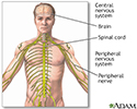
Central nervous system and peripheral nervous system - illustration
The central nervous system comprises the brain and spinal cord. The peripheral nervous system includes nerves outside the brain and spinal cord.
Central nervous system and peripheral nervous system
illustration
Review Date: 4/25/2022
Reviewed By: Joseph V. Campellone, MD, Department of Neurology, Cooper University Hospital, Camden, NJ. Review provided by VeriMed Healthcare Network. Also reviewed by David C. Dugdale, MD, Medical Director, Brenda Conaway, Editorial Director, and the A.D.A.M. Editorial team.


