Cranial mononeuropathy III - diabetic type
Diabetic third nerve palsy; Pupil-sparing third cranial nerve palsy; Ocular diabetic neuropathyThis diabetic type of cranial mononeuropathy III is a complication of diabetes. It causes double vision and eyelid drooping.
Diabetes
Diabetes is a long-term (chronic) disease in which the body cannot regulate the amount of sugar in the blood.

Eyelid drooping
Ptosis (eyelid drooping) in infants and children is when the upper eyelid is lower than it should be. This may occur in one or both eyes. Eyelid dr...

Causes
Mononeuropathy means that only one nerve is damaged. This disorder affects the third cranial nerve in the skull. This is one of the cranial nerves that control eye movement and the pupil of the eye.
Mononeuropathy
Mononeuropathy is damage to a single nerve, which results in loss of movement, sensation, or other function of that nerve.

This type of damage may occur along with diabetic peripheral neuropathy. Cranial mononeuropathy III is the most common cranial nerve disorder in people with diabetes. It is due to damage to the small blood vessels that feed the nerve.
Diabetic peripheral neuropathy
Nerve damage that occurs in people with diabetes is called diabetic neuropathy. This condition is a complication of diabetes.

Cranial mononeuropathy III can also occur in people who don't have diabetes.
Cranial mononeuropathy III
Cranial mononeuropathy III is a nerve disorder. It affects the function of the third cranial nerve. As a result, the person may have double vision ...

Symptoms
Symptoms may include:
- Double vision
Double vision
There are many types of eye problems and vision disturbances, such as: Halos Blurred vision (the loss of sharpness of vision and the inability to see...
 ImageRead Article Now Book Mark Article
 - Drooping of one eyelid (ptosis)
- Pain around the eye and forehead
- Change in size of the pupil (anisocoria)
Anisocoria
Anisocoria is unequal pupil size. The pupil is the black part in the center of the eye. It gets larger in dim light and smaller in bright light....
 ImageRead Article Now Book Mark Article
Neuropathy often develops within 7 days of onset of pain.
Exams and Tests
An exam of the eyes will determine whether only the third nerve is affected or if other nerves have also been damaged. Signs may include:
- Eyes that are not aligned
- Pupil reaction that is often normal
Your health care provider will do a complete exam to determine the possible effect on other parts of the nervous system. Depending on the suspected cause, you may need:
- Blood tests
- Tests to look at blood vessels in the brain (cerebral angiogram, CT angiogram, MR angiogram)
Cerebral angiogram
Cerebral angiography is a procedure that uses a special dye (contrast material) and x-rays to see how blood flows through the brain.
 ImageRead Article Now Book Mark Article
MR angiogram
Magnetic resonance angiography (MRA) is an MRI exam of the blood vessels. Unlike traditional angiography that involves placing a tube (catheter) int...
 ImageRead Article Now Book Mark Article
 - MRI or CT scan of the brain
MRI
A head MRI (magnetic resonance imaging) is an imaging test that uses powerful magnets and radio waves to create pictures of the brain and surrounding...
 ImageRead Article Now Book Mark Article
CT scan of the brain
A head computed tomography (CT) scan uses many x-rays to create pictures of the head, including the skull, brain, eye sockets, and sinuses.
 ImageRead Article Now Book Mark Article
 - Spinal tap (lumbar puncture)
Lumbar puncture
Cerebrospinal fluid (CSF) collection is a test to look at the fluid that surrounds the brain and spinal cord. CSF acts as a cushion, protecting the b...
 ImageRead Article Now Book Mark Article
You may need to be referred to a doctor who specializes in vision problems related to the nerves in the eye (neuro-ophthalmologist).
Treatment
There is no specific treatment to correct the nerve injury.
Treatments to help symptoms may include:
-
Close control of blood sugar level
Control of blood sugar level
When you have diabetes, you should have good control of your blood sugar (glucose). If your blood sugar is not controlled, serious health problems c...
 ImageRead Article Now Book Mark Article
 - Eye patch or glasses with prisms to reduce double vision
- Pain medicines
- Antiplatelet therapy
Antiplatelet therapy
Platelets are small particles in your blood that your body uses to form clots and stop bleeding. If you have too many platelets or your platelets st...
 ImageRead Article Now Book Mark Article
 - Surgery to correct eyelid drooping or eyes that are not aligned
Some people may recover without treatment.
Outlook (Prognosis)
The prognosis is good. Many people get better over 3 to 6 months. However, some people have permanent eye muscle weakness.
Possible Complications
Complications may include:
- Permanent eyelid drooping
- Permanent vision changes
When to Contact a Medical Professional
Contact your provider if you have double vision and it does not go away in a few minutes, especially if you also have eyelid drooping.
Prevention
Controlling your blood sugar level may reduce the risk of developing this disorder.
References
Brownlee M, Aiello LP, Sun JK, et al. Complications of diabetes mellitus. In: Melmed S, Auchus RJ, Goldfine AB, Rosen CJ, Kopp PA, eds. Williams Textbook of Endocrinology. 15th ed. Philadelphia, PA: Elsevier; 2025:chap 38.
Rucker JC, Seay MD. Cranial neuropathies. In: Jankovic J, Mazziotta JC, Pomeroy SL, Newman NJ, eds. Bradley and Daroff's Neurology in Clinical Practice. 8th ed. Philadelphia, PA: Elsevier; 2022:chap 103.
Tamhankar MA. Eye movement disorders: third, fourth, and sixth nerve palsies and other causes of diplopia and ocular misalignment. In: Liu GT, Volpe NJ, Galetta SL, eds. Liu, Volpe, and Galetta's Neuro-Ophthalmology. 3rd ed. Philadelphia, PA: Elsevier; 2019:chap 15.
Thurtell MJ, Rucker JC. Neuro-ophthalmology. In: Winn HR, ed. Youmans and Winn Neurological Surgery. 8th ed. Philadelphia, PA: Elsevier; 2023:chap 15.
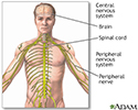
Central nervous system and peripheral nervous system - illustration
The central nervous system comprises the brain and spinal cord. The peripheral nervous system includes nerves outside the brain and spinal cord.
Central nervous system and peripheral nervous system
illustration
Review Date: 6/13/2024
Reviewed By: Joseph V. Campellone, MD, Department of Neurology, Cooper Medical School at Rowan University, Camden, NJ. Review provided by VeriMed Healthcare Network. Also reviewed by David C. Dugdale, MD, Medical Director, Brenda Conaway, Editorial Director, and the A.D.A.M. Editorial team.


