Malaria
Quartan malaria; Falciparum malaria; Biduoterian fever; Blackwater fever; Tertian malaria; PlasmodiumMalaria is a parasitic disease that involves high fevers, shaking chills, flu-like symptoms, and anemia.
Anemia
Anemia is a condition in which the body does not have enough healthy red blood cells. Red blood cells provide oxygen to body tissues. Different type...

Causes
Malaria is caused by a parasite. It is passed to humans by the bite of infected Anopheles mosquitoes. After infection, the parasites (called sporozoites) travel through the bloodstream to the liver. There, they mature and release another form of parasites, called merozoites. The parasites enter the bloodstream and infect red blood cells (RBCs).
The parasites multiply inside the red blood cells. The cells then break open within 48 to 72 hours and infect more red blood cells. The first symptoms usually occur 2 to 4 weeks after infection, though they can appear as early as 8 days or as long as a year after infection. The symptoms occur in cycles of 48 to 72 hours.
Most symptoms are caused by:
- The release of merozoites into the bloodstream
- Anemia resulting from the destruction of the red blood cells
- Large amounts of free hemoglobin being released into circulation after red blood cells break open, which can damage other organs such as the kidneys
Malaria can also be transmitted from a mother to her unborn baby (congenitally) and by blood transfusions. Malaria can be carried by mosquitoes in temperate climates, but the parasite disappears over the winter.
The disease is a major health problem in much of the tropics and subtropics. The Centers for Disease Control and Prevention reported that in 2020 there were about 241 million cases of malaria. About 627,000 people died of it. Malaria is a major disease hazard for travelers to warm climates.
In some areas of the world, mosquitoes that carry malaria have developed resistance to insecticides. In addition, the parasites have developed resistance to some antibiotics. These conditions have made it hard to control both the rate of infection and spread of this disease.
Symptoms
Symptoms include:
-
Anemia (condition in which the body doesn't have enough healthy red blood cells)
Anemia
Anemia is a condition in which the body does not have enough healthy red blood cells. Red blood cells provide oxygen to body tissues. Different type...
 ImageRead Article Now Book Mark Article
 -
Bloody stools
Bloody stools
Black or tarry stools with a foul smell are a sign of a problem in the upper digestive tract. It most often indicates that there is bleeding in the ...
 ImageRead Article Now Book Mark Article
 - Chills, fever, sweating
-
Coma
Coma
Decreased alertness is a state of reduced awareness and is often a serious condition. A coma is the most severe state of decreased alertness from whi...
Read Article Now Book Mark Article -
Convulsions
Convulsions
A seizure is the physical changes in behavior that occurs during an episode of specific types of abnormal electrical activity in the brain. The term ...
 ImageRead Article Now Book Mark Article
 - Headache
-
Jaundice
Jaundice
Jaundice is a yellow color of the skin, mucus membranes, or eyes. The yellow coloring comes from bilirubin, a byproduct of old red blood cells. Jau...
 ImageRead Article Now Book Mark Article
 - Muscle pain
- Nausea and vomiting
Exams and Tests
During a physical examination, the health care provider may find an enlarged liver or enlarged spleen.
Enlarged liver
Enlarged liver refers to swelling of the liver beyond its normal size. Hepatomegaly is another word to describe this problem. If both the liver and ...

Enlarged spleen
Splenomegaly is a larger-than-normal spleen. The spleen is an organ in the upper left part of the belly.

Tests that are done include:
- Rapid diagnostic tests, which are becoming more common because they are easier to use and require less training by laboratory technicians
- Malaria blood smears taken at 6 to 12 hour intervals to confirm the diagnosis
- A complete blood count (CBC) will identify anemia if it is present
Complete blood count
A complete blood count (CBC) test measures the following:The number of white blood cells (WBC count)The number of red blood cells (RBC count)The numb...
 ImageRead Article Now Book Mark Article
Treatment
Malaria, especially falciparum malaria, is a medical emergency that requires a hospital stay. Chloroquine is often used as an anti-malarial medicine. But chloroquine-resistant infections are common in some parts of the world.
Possible treatments for chloroquine-resistant infections include:
- Artemisinin derivative combinations, including artemether and lumefantrine
- Atovaquone-proguanil
- Quinine-based regimen, in combination with doxycycline or clindamycin
- Mefloquine, in combination with artesunate or doxycycline
The choice of medicine depends, in part, on where you got the infection.
Medical care, including fluids through a vein (IV) and other medicines and breathing (respiratory) support may be needed.
Outlook (Prognosis)
Outcome is expected to be good in most cases of malaria with treatment, but poor in falciparum infection with complications.
Possible Complications
Health problems that may result from malaria include:
- Brain infection (cerebritis)
- Destruction of blood cells (hemolytic anemia)
Hemolytic anemia
Anemia is a condition in which the body does not have enough healthy red blood cells. Red blood cells provide oxygen to body tissues. Normally, red ...
 ImageRead Article Now Book Mark Article
 -
Kidney failure
Kidney failure
Acute kidney failure is the rapid (less than 2 days) loss of your kidneys' ability to remove waste and help balance fluids and electrolytes in your b...
 ImageRead Article Now Book Mark Article
 - Liver failure
-
Meningitis
Meningitis
Meningitis is an infection of the membranes covering the brain and spinal cord. This covering is called the meninges.
 ImageRead Article Now Book Mark Article
 - Respiratory failure from fluid in the lungs (pulmonary edema)
Pulmonary edema
Pulmonary edema is an abnormal buildup of fluid in the lungs. This buildup of fluid leads to shortness of breath.
 ImageRead Article Now Book Mark Article
 - Rupture of the spleen leading to massive internal bleeding (hemorrhage)
When to Contact a Medical Professional
Contact your health care provider if you develop fever and headache after visiting any foreign country.
Prevention
Most people who live in areas where malaria is common have developed some immunity to the disease. Visitors will not have immunity and should take preventive medicines.
Immunity
The immune response is how your body recognizes and defends itself against bacteria, viruses, and substances that appear foreign and harmful....

It is important to see your provider well before your trip. This is because treatment may need to begin as long as 2 weeks before travel to the area, and continue for a month after you leave the area. Most travelers from the United States who contract malaria fail to take the right precautions.
The types of anti-malarial medicines prescribed depend on the area you visit. Travelers to South America, Africa, the Indian subcontinent, Asia, and the South Pacific should take one of the following medicines:
- Mefloquine
- Doxycycline
- Chloroquine
- Hydroxychloroquine
- Atovaquone-proguanil
Even pregnant women should consider taking preventive medicines because the risk to the fetus from the medicine is less than the risk of catching this infection.
Chloroquine has been the medicine of choice for protecting against malaria. But because of resistance, it is now only suggested for use in areas where Plasmodium vivax, P oval, and P malariae are present.
Falciparum malaria is becoming increasingly resistant to anti-malarial medicines. Recommended medicines include mefloquine, atovaquone/proguanil (Malarone), and doxycycline.
Prevent mosquito bites by:
- Wearing protective clothing over your arms and legs
- Using mosquito netting while sleeping
- Using insect repellent
For information on malaria and preventive medicines, visit the CDC website: www.cdc.gov/malaria/prevention/index.html.
References
Ansong D, Seydel KB, Taylor TE. Malaria. In: Ryan ET, Hill DR, Solomon T, Aronson NE, Endy TP, eds. Hunter's Tropical Medicine and Emerging Infectious Diseases. 10th ed. Philadelphia, PA: Elsevier; 2020:chap 101.
Fairhurst RM, Wellems TE. Malaria (Plasmodium species). In: Bennett JE, Dolin R, Blaser MJ, eds. Mandell, Douglas, and Bennett's Principles and Practice of Infectious Diseases. 9th ed. Philadelphia, PA: Elsevier; 2020:chap 274.
Freedman DO. Protection of travelers. In: Bennett JE, Dolin R, Blaser MJ, eds. Mandell, Douglas, and Bennett's Principles and Practice of Infectious Diseases. 9th ed. Philadelphia, PA: Elsevier; 2020:chap 318.
-
Malaria, microscopic view of cellular parasites - illustration
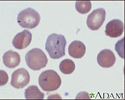
Malaria is a disease caused by parasites that are carried by mosquitoes. Once in the bloodstream, the parasite inhabits the red blood cell (RBC). This picture shows purple-stained malaria parasites inside red blood cells.
Malaria, microscopic view of cellular parasites
illustration
-
Mosquito, adult feeding on the skin - illustration
There are many different species of mosquito, which can carry some of the world's most common and significant infectious diseases, including West Nile, Malaria, yellow fever, viral encephalitis, and dengue fever. (Image courtesy of the Centers for Disease Control and Prevention.)
Mosquito, adult feeding on the skin
illustration
-
Mosquito, egg raft - illustration
Mosquitoes of the Culex species lay their eggs in the form of egg rafts that float in still or stagnant water. The mosquito lays the eggs one at a time sticking them together in the shape of a raft. An egg raft can contain from 100 to 400 eggs. The eggs go through larval and pupal stages and feed on micro-organisms before developing into flying mosquitoes. (Image courtesy of the Centers for Disease Control and Prevention.)
Mosquito, egg raft
illustration
-
Mosquito, larvae - illustration
This picture shows mosquito larvae, an early stage of the mosquito life cycle. (Image courtesy of the Centers for Disease Control and Prevention.)
Mosquito, larvae
illustration
-
Mosquito, pupa - illustration
These are mosquito pupa. This is another stage in the development of the mosquito. (Image courtesy of the Centers for Disease Control and Prevention.)
Mosquito, pupa
illustration
-
Malaria, microscopic view of cellular parasites - illustration
Malarial parasites are visible within the red blood cells. They are stained a dark bluish color.
Malaria, microscopic view of cellular parasites
illustration
-
Malaria, photomicrograph of cellular parasites - illustration
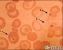
Malaria is a disease caused by parasites. This picture shows dark orange-stained malaria parasites inside red blood cells (a) and outside the cells (b). Note the large cells that look like targets; it is unknown how these target cells are related to this disease.
Malaria, photomicrograph of cellular parasites
illustration
-
Malaria - illustration
Malaria is caused by a parasite transmitted from one human to another via the bite of an infected Anopheles mosquito. The parasites migrate to the liver, mature and enter the bloodstream, where they rupture red blood cells. An infected pregnant woman can transmit malaria to her unborn child.
Malaria
illustration
-
Digestive system organs - illustration
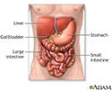
The digestive system organs in the abdominal cavity include the liver, gallbladder, stomach, small intestine and large intestine.
Digestive system organs
illustration
-
Malaria, microscopic view of cellular parasites - illustration
Malaria is a disease caused by parasites that are carried by mosquitoes. Once in the bloodstream, the parasite inhabits the red blood cell (RBC). This picture shows purple-stained malaria parasites inside red blood cells.
Malaria, microscopic view of cellular parasites
illustration
-
Mosquito, adult feeding on the skin - illustration
There are many different species of mosquito, which can carry some of the world's most common and significant infectious diseases, including West Nile, Malaria, yellow fever, viral encephalitis, and dengue fever. (Image courtesy of the Centers for Disease Control and Prevention.)
Mosquito, adult feeding on the skin
illustration
-
Mosquito, egg raft - illustration
Mosquitoes of the Culex species lay their eggs in the form of egg rafts that float in still or stagnant water. The mosquito lays the eggs one at a time sticking them together in the shape of a raft. An egg raft can contain from 100 to 400 eggs. The eggs go through larval and pupal stages and feed on micro-organisms before developing into flying mosquitoes. (Image courtesy of the Centers for Disease Control and Prevention.)
Mosquito, egg raft
illustration
-
Mosquito, larvae - illustration
This picture shows mosquito larvae, an early stage of the mosquito life cycle. (Image courtesy of the Centers for Disease Control and Prevention.)
Mosquito, larvae
illustration
-
Mosquito, pupa - illustration
These are mosquito pupa. This is another stage in the development of the mosquito. (Image courtesy of the Centers for Disease Control and Prevention.)
Mosquito, pupa
illustration
-
Malaria, microscopic view of cellular parasites - illustration
Malarial parasites are visible within the red blood cells. They are stained a dark bluish color.
Malaria, microscopic view of cellular parasites
illustration
-
Malaria, photomicrograph of cellular parasites - illustration
Malaria is a disease caused by parasites. This picture shows dark orange-stained malaria parasites inside red blood cells (a) and outside the cells (b). Note the large cells that look like targets; it is unknown how these target cells are related to this disease.
Malaria, photomicrograph of cellular parasites
illustration
-
Malaria - illustration
Malaria is caused by a parasite transmitted from one human to another via the bite of an infected Anopheles mosquito. The parasites migrate to the liver, mature and enter the bloodstream, where they rupture red blood cells. An infected pregnant woman can transmit malaria to her unborn child.
Malaria
illustration
-
Digestive system organs - illustration
The digestive system organs in the abdominal cavity include the liver, gallbladder, stomach, small intestine and large intestine.
Digestive system organs
illustration
Review Date: 5/19/2023
Reviewed By: Jatin M. Vyas, MD, PhD, Associate Professor in Medicine, Harvard Medical School; Associate in Medicine, Division of Infectious Disease, Department of Medicine, Massachusetts General Hospital, Boston, MA. Also reviewed by David C. Dugdale, MD, Medical Director, Brenda Conaway, Editorial Director, and the A.D.A.M. Editorial team.


