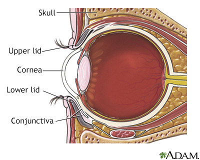
Dry eye syndrome
You need tears to moisten your eyes and to wash away particles that have gotten into your eyes. A healthy tear film on the eye is necessary for good vision.
Dry eyes develop when the eye is unable to maintain a healthy coating of tears.
Causes
Dry eye syndrome commonly occurs in people who are otherwise healthy. It becomes more common with older age. This can occur due to hormonal changes that make your eyes produce fewer tears. Dry eye syndrome is sometimes caused or worsened by a condition called meibomianitis, which changes the normal tear film.
Meibomianitis
Meibomianitis is inflammation of the meibomian glands, a group of oil-releasing (sebaceous) glands in the eyelids. These glands have tiny openings t...

Other common causes of dry eyes include:
- Dry environment or workplace (wind, air conditioning)
- Sun exposure
- Smoking or second-hand smoke exposure
- Cold or allergy medicines
- Wearing contact lenses
Dry eye syndrome can also be caused by:
- Heat or chemical burns
- Previous eye surgery
- Use of eye drops for other eye diseases
- An uncommon autoimmune disorder in which the glands that produce tears are destroyed (Sjögren syndrome)
Autoimmune disorder
An autoimmune disorder occurs when the body's immune system attacks and destroys healthy body tissue by mistake. There are more than 80 autoimmune d...
 ImageRead Article Now Book Mark Article
Sjögren syndrome
Sjögren syndrome is an autoimmune disorder in which the glands that produce tears and saliva are destroyed. This causes dry mouth and dry eyes. The...
 ImageRead Article Now Book Mark Article
Symptoms
Symptoms may include:
- Blurred vision
Blurred vision
There are many types of eye problems and vision disturbances, such as: Halos Blurred vision (the loss of sharpness of vision and the inability to see...
 ImageRead Article Now Book Mark Article
 - Burning, itching, or redness in the eye
- Gritty or scratchy feeling in the eye
- Sensitivity to light
Exams and Tests
Tests may include:
- Visual acuity measurement
- Slit lamp exam
- Diagnostic staining of the cornea and tear film
- Measurement of tear film break-up time (TBUT)
- Measurement of rate of tear production (Schirmer test)
- Measurement of concentration of tears (osmolality)
Treatment
The first step in treatment is artificial tears. These come as preserved (screw cap bottle) and unpreserved (twist open vial or multidose bottle). Preserved tears are more convenient, but some people are sensitive to preservatives. There are many brands available without a prescription.
Start using the drops at least 2 to 4 times per day. If your symptoms are not better after a couple of weeks of regular use:
- Increase use (up to every 2 hours if using preservative free drops).
- Change to unpreserved drops if you have been using the preserved type.
- Try a different brand.
- Talk to your health care provider if you cannot find a brand that works for you.
Other treatments for dry eyes may include:
- Fish oil 2 to 3 times per day
- Glasses, goggles or contact lenses that keep moisture in the eyes
- Medicines such as cyclosporine (Restasis) or lifitegrast (Xiidra), corticosteroid eye drops, and oral tetracycline and doxycycline
- Tiny plugs placed in the tear drainage ducts to help moisture stay on the surface of the eye longer
Other helpful steps include:
- DO NOT smoke.
- Avoid second-hand smoke, direct wind, and air conditioning.
- Use a humidifier, particularly in the winter.
- Limit allergy and cold medicines that may dry you out and worsen your symptoms.
- Purposefully blink more often. Rest your eyes once in a while.
- Clean eyelashes regularly and apply warm compresses.
Some dry eye symptoms are due to sleeping with the eyes slightly open. Lubricating ointments work best for this problem. You should use them only in small amounts since they can blur your vision. It is best to use them before sleep.
Surgery may be helpful if symptoms are because the eyelids are in an abnormal position.
Outlook (Prognosis)
Most people with dry eye syndrome have only discomfort, and no vision loss.
Possible Complications
In severe cases, the clear covering on the eye (cornea) may become damaged or infected.
When to Contact a Medical Professional
Contact your provider right away if:
- You have red or painful eyes.
- You have flaking, discharge, or a sore on your eye or eyelid.
- You have had an injury to your eye, or if you have a bulging eye or a drooping eyelid.
- You have joint pain, swelling, or stiffness and a dry mouth along with dry eye symptoms.
- Your eyes do not get better with self-care within a few days.
Prevention
Stay away from dry environments and things that irritate your eyes to help prevent symptoms.
Reviewed By
Audrey Tai, DO, MS, Athena Eye Care, Mission Viejo, CA. Also reviewed by David C. Dugdale, MD, Medical Director, Brenda Conaway, Editorial Director, and the A.D.A.M. Editorial team.
Brissette AR, Bohm KJ, Starr CE. Dry eye overview: classification and treatment overview. In: Mannis MJ, Holland EJ, eds. Cornea. 5th ed. Philadelphia, PA: Elsevier; 2022:chap 31.
Dhawlikar NS, Holdstein MH, Rao NK. Dry eye disease. In: Yanoff M, Duker JS, eds. Ophthalmology. 6th ed. Philadelphia, PA: Elsevier; 2023:chap 4.23
Jeang LA. Dry eye syndrome. In: Kellerman RD, Rakel DP, Heidelbaugh JJ, Lee EM, eds. Conn's Current Therapy 2024. Philadelphia, PA: Elsevier; 2024:527-529.


 All rights reserved.