
Thyroid cancer - papillary carcinoma
Papillary carcinoma of the thyroid; Papillary thyroid cancer; Papillary thyroid carcinomaPapillary carcinoma of the thyroid is the most common cancer of the thyroid gland. The thyroid gland is located in front of the lower neck.
Causes
About 85% of all thyroid cancers diagnosed in the United States are the papillary carcinoma type. It is more common in women than in men. It may occur in childhood, but is most often seen in adults between ages 20 and 60.
The cause of this cancer is not known. A genetic defect or family history of the disease may be a risk factor.
Radiation increases the risk for developing thyroid cancer. Exposure may occur from:
- High-dose external radiation treatments to the neck, especially during childhood, used to treat childhood cancer or some noncancerous childhood conditions
- Radiation exposure from nuclear plant disasters
Radiation given through a vein (through an IV) during medical tests and treatments does not increase the risk for developing thyroid cancer.
Symptoms
Thyroid cancer often begins as a small lump (nodule) in the thyroid gland.
While some small lumps may be cancer, most (90%) thyroid nodules are harmless and are not cancerous.
Most of the time, there are no other symptoms.
Exams and Tests
If you have a lump on your thyroid, your health care provider may order the following tests:
- Blood tests.
- Ultrasound of the thyroid gland and neck region.
Thyroid gland and neck region
A thyroid ultrasound is an imaging method to see the thyroid, a gland in the neck that regulates metabolism (the many processes that control the rate...
 ImageRead Article Now Book Mark Article
 -
CT scan of the neck or MRI to determine the size of the tumor.
CT scan
A computed tomography (CT) scan is an imaging method that uses x-rays to create pictures of cross-sections of the body. Related tests include:Abdomin...
 ImageRead Article Now Book Mark Article
MRI
A magnetic resonance imaging (MRI) scan is an imaging test that uses powerful magnets and radio waves to create pictures of the body. It does not us...
 ImageRead Article Now Book Mark Article
 - An examination of the airway with a fiberoptic scope (laryngoscopy) may show a paralyzed vocal cord.
Laryngoscopy
Laryngoscopy is an exam of the back of your throat, including your voice box (larynx). Your voice box contains your vocal cords and allows you to sp...
 ImageRead Article Now Book Mark Article
 -
Fine needle aspiration biopsy (FNAB) to determine if the lump is cancerous. FNAB may be performed if the ultrasound shows that the lump is less than 1 centimeter.
Fine needle aspiration biopsy (FNAB)
Fine needle aspiration of the thyroid gland is a procedure to remove thyroid cells for examination. The thyroid gland is a butterfly-shaped gland lo...
 ImageRead Article Now Book Mark Article
Genetic testing may be done on the biopsy sample to see what genetic changes (mutations) may be present. Knowing this may help guide treatment recommendations.
Genetic testing
The genes in our cells play important roles. They affect hair and eye color and other traits passed on from parent to child. Genes also tell cells ...
Thyroid function tests are often normal in people with thyroid cancer.
Thyroid function tests
Thyroid function tests are used to check whether your thyroid is working normally. The most common thyroid function tests are:Free T4 (free thyroxine...

Treatment
Thyroid cancer treatment may include:
-
Surgery
Surgery
Thyroid gland removal is surgery to remove all or part of the thyroid gland. The thyroid gland is a butterfly-shaped gland located inside the front ...
 ImageRead Article Now Book Mark Article
 -
Radioactive iodine therapy
Radioactive iodine therapy
Radioiodine therapy uses radioactive iodine to kill thyroid cells and shrink the thyroid gland. It is used to treat certain diseases of the thyroid ...
 ImageRead Article Now Book Mark Article
 - Thyroid suppression therapy (thyroid hormone replacement therapy)
- External beam radiation therapy (EBRT)
Surgery is done to remove as much of the cancer as possible. The bigger the lump, the more of the thyroid gland must be removed. Often, the entire gland is taken out.
After the surgery, you may receive radioiodine therapy, which is often taken by mouth. This substance kills any remaining thyroid tissue. It also helps make medical images clearer, so doctors can see if there is any cancer left behind or if it comes back later.
Further management of your cancer will depend on many factors such as:
- Size of any tumor present
- Location of the tumor
- Growth rate of the tumor
- Symptoms you may have
- Your own preferences
If surgery is not an option, external radiation therapy can be useful.
After surgery or radioiodine therapy, you will need to take medicine called levothyroxine for the rest of your life. This replaces the hormone the thyroid would normally make.
Your provider will likely have you take a blood test every several months to check thyroid hormone levels. Other follow-up tests that may be done after treatment for thyroid cancer include:
- Ultrasound of the thyroid
- An imaging test called a radioactive iodine (I-131) uptake scan
Radioactive iodine (I-131) uptake scan
Radioactive iodine uptake (RAIU) tests thyroid function. It measures how much radioactive iodine is taken up by your thyroid gland in a certain time...
 ImageRead Article Now Book Mark Article
 - Repeat FNAB
Support Groups
You can ease the stress of illness by joining a cancer support group. Sharing with others who have common experiences and problems can help you not feel alone.
Cancer support group
The following organizations are good resources for information on cancer:American Cancer Society. Support and online communities. www. cancer. org/...
Outlook (Prognosis)
The survival rate for papillary thyroid cancer is excellent. More than 90% of adults with this cancer survive at least 10 to 20 years. The prognosis is better for people who are younger than 40 and for those with smaller tumors.
The following factors may decrease the survival rate:
- Older than 55 years of age
- Cancer that has spread to distant parts of the body
- Cancer that has spread to soft tissue around the thyroid
- Large tumor
Possible Complications
Complications include:
- Accidental removal of the parathyroid glands, which help regulate blood calcium levels
- Damage to the nerve that controls the vocal cords
- Spreading of cancer to lymph nodes (rare)
- Spreading of cancer to other sites (metastasis)
Metastasis
Metastasis is the movement or spreading of cancer cells from one organ or tissue to another. Cancer cells usually spread through the blood or the ly...
 ImageRead Article Now Book Mark Article
When to Contact a Medical Professional
Contact your provider if you have a lump in your neck.
References
NCCN Clinical Practice Guidelines in Oncology NCCN Guidelines) website. Thyroid carcinoma, Version 2.2024 - March 12, 2024. www.nccn.org/professionals/physician_gls/pdf/thyroid.pdf. Accessed May 10, 2024.
National Cancer Institute website. Thyroid cancer treatment (PDQ) - health provisional version. www.cancer.gov/cancertopics/pdq/treatment/thyroid/HealthProfessional. Updated April 11, 2024. Accessed May 3, 2024.
Pearce EN, Hollenberg AN. Thyroid. In: Goldman L, Cooney KA, eds. Goldman-Cecil Medicine. 27th ed. Philadelphia, PA: Elsevier; 2024:chap 207.
Sipos JA, Haugen BR. Papillary thyroid cancer. In: Randolph GW, ed. Surgery of the thyroid and parathyroid glands. 3rd ed. Philadelphia, PA: Elsevier; 2021:chap. 19.
Thompson LDR. Malignant neoplasms of the thyroid gland. In: Thompson LDR, Bishop JA, eds. Head and Neck Pathology. 3rd ed. Philadelphia, PA: Elsevier; 2019:chap 25.
-
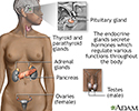
Endocrine glands - illustration
Endocrine glands release hormones (chemical messengers) into the bloodstream to be transported to various organs and tissues throughout the body. For instance, the pancreas secretes insulin, which allows the body to regulate levels of sugar in the blood. The thyroid gets instructions from the pituitary to secrete hormones which determine the rate of metabolism in the body (the more hormone in the bloodstream, the faster the chemical activity; the less hormone, the slower the activity).
Endocrine glands
illustration
-
Thyroid cancer - CT scan - illustration
This CT scan of the upper chest (thorax) shows a malignant thyroid tumor (cancer). The dark area around the trachea (marked by the white U-shaped tip of the respiratory tube) is an area where normal tissue has been eroded and died (necrosis) as a result of tumor growth.
Thyroid cancer - CT scan
illustration
-
Thyroid cancer - CT scan - illustration
This CT scan shows a thyroid cancer tumor in the throat, encircling, narrowing, and displacing the windpipe (trachea).
Thyroid cancer - CT scan
illustration
-
Thyroid enlargement - scintiscan - illustration
This image shows the enlargement of the thyroid gland and extension down behind the breastbone (retrosternal space). The image, called a scintiscan, was generated using a radioactive isotope.
Thyroid enlargement - scintiscan
illustration
-
Thyroid gland - illustration
The thyroid gland, a part of the endocrine (hormone) system, plays a major role in regulating the body's metabolism.
Thyroid gland
illustration


-
Endocrine glands - illustration
Endocrine glands release hormones (chemical messengers) into the bloodstream to be transported to various organs and tissues throughout the body. For instance, the pancreas secretes insulin, which allows the body to regulate levels of sugar in the blood. The thyroid gets instructions from the pituitary to secrete hormones which determine the rate of metabolism in the body (the more hormone in the bloodstream, the faster the chemical activity; the less hormone, the slower the activity).
Endocrine glands
illustration
-
Thyroid cancer - CT scan - illustration
This CT scan of the upper chest (thorax) shows a malignant thyroid tumor (cancer). The dark area around the trachea (marked by the white U-shaped tip of the respiratory tube) is an area where normal tissue has been eroded and died (necrosis) as a result of tumor growth.
Thyroid cancer - CT scan
illustration
-
Thyroid cancer - CT scan - illustration
This CT scan shows a thyroid cancer tumor in the throat, encircling, narrowing, and displacing the windpipe (trachea).
Thyroid cancer - CT scan
illustration
-
Thyroid enlargement - scintiscan - illustration
This image shows the enlargement of the thyroid gland and extension down behind the breastbone (retrosternal space). The image, called a scintiscan, was generated using a radioactive isotope.
Thyroid enlargement - scintiscan
illustration
-
Thyroid gland - illustration
The thyroid gland, a part of the endocrine (hormone) system, plays a major role in regulating the body's metabolism.
Thyroid gland
illustration
Review Date: 3/31/2024
Reviewed By: Todd Gersten, MD, Hematology/Oncology, Florida Cancer Specialists & Research Institute, Wellington, FL. Review provided by VeriMed Healthcare Network. Also reviewed by David C. Dugdale, MD, Medical Director, Brenda Conaway, Editorial Director, and the A.D.A.M. Editorial team.
 All rights reserved.