Hepatic hemangioma
Liver hemangioma; Hemangioma of the liver; Cavernous hepatic hemangioma; Infantile hemangioendothelioma; Multinodular hepatic hemangiomatosisA hepatic hemangioma is a liver mass made of widened (dilated) blood vessels. It is not cancerous.
Causes
A hepatic hemangioma is the most common type of liver mass that is not caused by cancer. It may be a birth defect.
Hepatic hemangiomas can occur at any time. They are most common in people in their 30s to 50s. Women get these masses more often than men. The masses are often bigger in size.
Babies may develop a type of hepatic hemangioma called benign infantile hemangioendothelioma. This is also known as multinodular hepatic hemangiomatosis. This is a rare, noncancerous tumor that has been linked to high rates of heart failure and death in infants. Infants are most often diagnosed by the time they are 6 months old.
Tumor
A tumor is an abnormal growth of body tissue. Tumors can be cancerous (malignant) or noncancerous (benign).
Read Article Now Book Mark ArticleSymptoms
Some hemangiomas may cause bleeding or interfere with organ function. Most do not produce symptoms. In rare cases, the hemangioma may rupture.
Exams and Tests
In most cases, the condition is not found until liver images are taken for some other reason. If the hemangioma ruptures, the only sign may be an enlarged liver.
Babies with benign infantile hemangioendothelioma may have:
- A growth in the abdomen
- Anemia
Anemia
Anemia is a condition in which the body does not have enough healthy red blood cells. Red blood cells provide oxygen to body tissues. Different type...
 ImageRead Article Now Book Mark Article
 - Signs of heart failure
Heart failure
Heart failure is a condition in which the heart is no longer able to pump oxygen-rich blood to the rest of the body efficiently. This causes symptom...
 ImageRead Article Now Book Mark Article
The following tests may be performed:
- Blood tests
- CT scan of the liver
CT scan of the liver
An abdominal CT scan is an imaging test that uses x-rays to create cross-sectional pictures of the belly area. CT stands for computed tomography....
 ImageRead Article Now Book Mark Article
 - Hepatic angiogram
- MRI
MRI
An abdominal magnetic resonance imaging scan is an imaging test that uses powerful magnets and radio waves. The waves create pictures of the inside ...
 ImageRead Article Now Book Mark Article
 - Single-photon emission computed tomography (SPECT)
- Ultrasound of the abdomen
Ultrasound of the abdomen
Abdominal ultrasound is a type of imaging test. It is used to look at organs in the abdomen, including the liver, gallbladder, pancreas, and kidneys...
 ImageRead Article Now Book Mark Article
Treatment
Most of these tumors are treated only if there is ongoing pain.
Treatment for infantile hemangioendothelioma depends on the child's growth and development. The following treatments may be needed:
- Inserting a material in a blood vessel of the liver to block it (embolization)
- Tying off (ligating) a liver artery
- Medicines for heart failure
- Surgery to remove the tumor
Outlook (Prognosis)
Surgery can cure a tumor in an infant if it is only in one lobe of the liver. This can be done even if the child has heart failure.
Possible Complications
Pregnancy and estrogen-based medicines can cause these tumors to grow.
The tumor may rupture in rare cases.
References
Cameron J. Liver: management of liver hemangiomas. In: Cameron J, ed. Current Surgical Therapy. 14th ed. Philadelphia, PA: Elsevier; 2023:351-412.
Coleman DM, Mendes BC, Tollefson MM, Bower TC. Pediatric vascular tumors. In: Sidawy AN, Perler BA, eds. Rutherford's Vascular Surgery and Endovascular Therapy. 10th ed. Philadelphia, PA: Elsevier; 2023:chap 187.
Di Bisceglie AM, Befeler AS. Hepatic tumors and cysts. In: Feldman M, Friedman LS, Brandt LJ, eds. Sleisenger and Fordtran's Gastrointestinal and Liver Disease: Pathophysiology/Diagnosis/Management. 11th ed. Philadelphia, PA: Elsevier; 2021:chap 96.
Hemangioma - angiogram - illustration
This angiogram (an X-ray taken after dye has been injected into the blood stream) shows a mass of blood vessels (hemangioma) in the liver.
Hemangioma - angiogram
illustration
Hemangioma - CT scan - illustration
This upper abdominal CT scan shows a blood vessel tumor (hemangioma) in the liver.
Hemangioma - CT scan
illustration
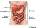
Digestive system organs - illustration
The digestive system organs in the abdominal cavity include the liver, gallbladder, stomach, small intestine and large intestine.
Digestive system organs
illustration
Review Date: 5/2/2023
Reviewed By: Michael M. Phillips, MD, Emeritus Professor of Medicine, The George Washington University School of Medicine, Washington, DC. Also reviewed by David C. Dugdale, MD, Medical Director, Brenda Conaway, Editorial Director, and the A.D.A.M. Editorial team.


