Hepatic vein obstruction (Budd-Chiari)
Hepatic vein obstruction is a blockage of the hepatic vein, which carries blood away from the liver.
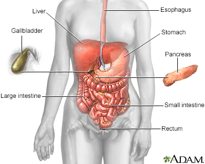
Hepatic
The term "hepatic" refers to the liver. For example, the hepatic duct drains bile from the liver.
Read Article Now Book Mark ArticleCauses
Hepatic vein obstruction prevents blood from flowing out of the liver and back to the heart. This blockage can cause liver damage. Obstruction of this vein can be caused by a tumor or growth pressing on the vessel, or by a clot in the vessel (hepatic vein thrombosis).
Tumor
A tumor is an abnormal growth of body tissue. Tumors can be cancerous (malignant) or noncancerous (benign).
Read Article Now Book Mark ArticleMost often, it is caused by conditions that make blood clots more likely to form, including:
- Abnormal growth of cells in the bone marrow (myeloproliferative disorders)
- Cancers
- Chronic inflammatory or autoimmune diseases
Autoimmune
An autoimmune disorder occurs when the body's immune system attacks and destroys healthy body tissue by mistake. There are more than 80 autoimmune d...
 ImageRead Article Now Book Mark Article
 - Infections
- Inherited (hereditary) or acquired problems with excessive blood clotting
- Oral contraceptives
- Pregnancy
Hepatic vein blockage is the most common cause of Budd-Chiari syndrome.
Symptoms
Symptoms include:
- Abdominal swelling or stretching due to fluid in the abdomen
- Pain in the right upper abdomen
- Vomiting blood
- Yellowing of the skin (jaundice)
Exams and Tests
One of the signs is swelling of the abdomen from fluid buildup (ascites). The liver is often swollen and tender.
Ascites
Ascites is the build-up of fluid in the space between the lining of the abdomen and abdominal organs.

Tests include:
-
CT scan or MRI of the abdomen
CT scan
A computed tomography (CT) scan is an imaging method that uses x-rays to create pictures of cross-sections of the body. Related tests include:Abdomin...
 ImageRead Article Now Book Mark Article
MRI of the abdomen
An abdominal magnetic resonance imaging scan is an imaging test that uses powerful magnets and radio waves. The waves create pictures of the inside ...
 ImageRead Article Now Book Mark Article
 - Doppler ultrasound of the liver veins
-
Liver biopsy
Liver biopsy
A liver biopsy is a test that takes a sample of tissue from the liver for examination.
 ImageRead Article Now Book Mark Article
 -
Liver function tests
Liver function tests
Liver function tests are common tests that are used to see how well the liver is working. Tests include:AlbuminAlpha-1 antitrypsinAlkaline phosphata...
 ImageRead Article Now Book Mark Article
 -
Ultrasound of the liver
Ultrasound
Ultrasound uses high-frequency sound waves to make images of organs and structures inside the body.
 ImageRead Article Now Book Mark Article
Treatment
Treatment varies, depending on the cause of the blockage.
Your health care provider may recommend the following medicines:
- Blood thinners (anticoagulants)
- Clot-busting medicines (thrombolytic treatment)
- Medicines to treat the liver disease, including ascites
Surgery may be recommended. This may involve:
-
Angioplasty and stent placement
Angioplasty
The blood vessels that bring blood to your brain and face are called the carotid arteries. You have a carotid artery on each side of your neck. The...
 ImageRead Article Now Book Mark Article
Stent
A stent is a tiny tube placed into a hollow structure in your body. This structure can be an artery, a vein, or another structure, such as the tube ...
 ImageRead Article Now Book Mark Article
 -
Transjugular intrahepatic portosystemic shunt (TIPS)
Transjugular intrahepatic portosystemic...
Transjugular intrahepatic portosystemic shunt (TIPS) is a procedure to create new connections between two blood vessels in your liver. You may need ...
 ImageRead Article Now Book Mark Article
 - Venous shunt surgery
- Liver transplant
Possible Complications
Hepatic vein obstruction can get worse and lead to cirrhosis and liver failure. This can be life threatening.
Cirrhosis
Cirrhosis is scarring of the liver and poor liver function. It is the last stage of chronic liver disease.

When to Contact a Medical Professional
Contact your provider if:
- You have symptoms of hepatic vein obstruction
- You are being treated for this condition and you develop new symptoms
Reviewed By
Jenifer K. Lehrer, MD, Department of Gastroenterology, Aria - Jefferson Health Torresdale, Jefferson Digestive Diseases Network, Philadelphia, PA. Review provided by VeriMed Healthcare Network. Also reviewed by David C. Dugdale, MD, Medical Director, Brenda Conaway, Editorial Director, and the A.D.A.M. Editorial team.
Kahi CJ. Vascular diseases of the gastrointestinal tract. In: Goldman L, Cooney KA, eds. Goldman-Cecil Medicine. 27th ed. Philadelphia, PA: Elsevier; 2024:chap 129.
Nery FG, Valla DC. Vascular diseases of the liver. In: Feldman M, Friedman LS, Brandt LJ, eds. Sleisenger and Fordtran's Gastrointestinal and Liver Disease. 11th ed. Philadelphia, PA: Elsevier; 2021:chap 85.
Disclaimer


 All rights reserved.