
Myocardial contusion
Blunt myocardial injuryMyocardial contusion is a bruise of the heart muscle.
Causes
The most common causes are:
- Car crashes
- Getting hit by a car
-
Cardiopulmonary resuscitation (CPR)
Cardiopulmonary resuscitation
CPR stands for cardiopulmonary resuscitation. It is an emergency life-saving procedure that is done when someone's breathing or heartbeat has stoppe...
Read Article Now Book Mark Article - Falling from a height, most often greater than 20 feet (6 meters)
Symptoms
A severe myocardial contusion may lead to signs and symptoms of a heart attack.
Symptoms can include:
- Pain in the front of the ribs or breastbone
- Feeling that your heart is racing
- Lightheadedness
- Nausea or vomiting
- Shortness of breath
- Weakness
Exams and Tests
The health care provider will perform a physical exam. This may show:
- Bruise or scrapes on the chest wall
- Crunching sensation when touching the skin if there are rib fractures and puncture of the lung
- Fast heartbeat
-
Irregular heartbeat
Irregular heartbeat
Palpitations are feelings or sensations that your heart is pounding or racing. They can be felt in your chest, throat, or neck. You may:Have an unpl...
 ImageRead Article Now Book Mark Article
 -
Low blood pressure
Low blood pressure
Low blood pressure occurs when blood pressure is below normal. This means the heart, brain, and other parts of the body may not get enough blood. I...
 ImageRead Article Now Book Mark Article
 - Rapid or shallow breathing
- Tenderness to the touch
- Abnormal chest wall movement from rib fractures
Tests may include:
- Blood tests (cardiac enzymes, such as Troponin-I or T or CKMB)
-
Chest x-ray
Chest x-ray
A chest x-ray is an x-ray of the chest, lungs, heart, large arteries, ribs, and diaphragm.
 ImageRead Article Now Book Mark Article
 - CT scan of the chest
-
Electrocardiogram (ECG)
Electrocardiogram
An electrocardiogram (ECG) is a test that records the electrical activity of the heart.
 ImageRead Article Now Book Mark Article
 -
Echocardiogram
Echocardiogram
An echocardiogram is a test that uses sound waves to create pictures of the heart. The picture and information it produces is more detailed than a s...
 ImageRead Article Now Book Mark Article
These tests may show:
- Problems with the heart wall and the ability for the heart to contract
- Fluid or blood in the thin sac surrounding the heart (pericardium)
- Rib fractures, lung or blood vessel injury
- Problem with the heart's electrical signaling (such as a bundle branch block or other heart block)
- Fast heartbeat starting at the sinus node of the heart (sinus tachycardia)
Tachycardia
An arrhythmia is a disorder of the heart rate (pulse) or heart rhythm. The heart can beat too fast (tachycardia), too slow (bradycardia), or irregul...
 ImageRead Article Now Book Mark Article
 - Abnormal heartbeat starting in the ventricles or lower chambers of the heart (ventricular dysrhythmia)
Treatment
In most cases, you will be closely monitored for at least 24 hours. An ECG will be done continually to check your heart beat and function.
Emergency room treatment may include:
- Catheter placement through a vein (IV)
- Medicines to relieve pain, heart rate disturbances, or low blood pressure
- Pacemaker (temporary, may be permanent later)
- Oxygen
Other therapies may be used to treat a heart injury, include:
- Chest tube placement
- Draining blood from around the heart
- Surgery to repair blood vessels in the chest
Outlook (Prognosis)
People with a mild myocardial contusion will recover completely most of the time.
Serious heart injuries can increase your risk for heart failure or heart rhythm problems.
Prevention
The following safety tips may help prevent a heart bruise:
- Wear a seat belt when driving.
- Choose a car with air bags.
- Take steps to ensure safety when working at heights.
References
Boccalandro F, Shreyder K. Traumatic heart disease. In: Levine GN, ed. Cardiology Secrets. 6th ed. Philadelphia, PA: Elsevier; 2023:chap 72.
Cameron J. Trauma and emergency care. In: Cameron J, ed. Current Surgical Therapy. 14th ed. Philadelphia, PA: Elsevier; 2023:chap 17.
Raja AS. Thoracic trauma. In: Walls RM, ed. Rosen's Emergency Medicine: Concepts and Clinical Practice. 10th ed. Philadelphia, PA: Elsevier; 2023:chap 37.
-
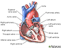
Heart - section through the middle - illustration
The interior of the heart is composed of valves, chambers, and associated vessels.
Heart - section through the middle
illustration
-
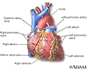
Heart - front view - illustration
The external structures of the heart include the ventricles, atria, arteries and veins. Arteries carry blood away from the heart while veins carry blood into the heart. The vessels colored blue indicate the transport of blood with relatively low content of oxygen and high content of carbon dioxide. The vessels colored red indicate the transport of blood with relatively high content of oxygen and low content of carbon dioxide.
Heart - front view
illustration
-
Heart - section through the middle - illustration
The interior of the heart is composed of valves, chambers, and associated vessels.
Heart - section through the middle
illustration
-
Heart - front view - illustration
The external structures of the heart include the ventricles, atria, arteries and veins. Arteries carry blood away from the heart while veins carry blood into the heart. The vessels colored blue indicate the transport of blood with relatively low content of oxygen and high content of carbon dioxide. The vessels colored red indicate the transport of blood with relatively high content of oxygen and low content of carbon dioxide.
Heart - front view
illustration
Review Date: 5/8/2024
Reviewed By: Thomas S. Metkus, MD, Assistant Professor of Medicine and Surgery, Johns Hopkins University School of Medicine, Baltimore, MD. Also reviewed by David C. Dugdale, MD, Medical Director, Brenda Conaway, Editorial Director, and the A.D.A.M. Editorial team.
 All rights reserved.