

Hypertrophic cardiomyopathy
Cardiomyopathy - hypertrophic (HCM); IHSS; Idiopathic hypertrophic subaortic stenosis; Asymmetric septal hypertrophy; ASH; HOCM; Hypertrophic obstructive cardiomyopathyHypertrophic cardiomyopathy (HCM) is a condition in which the heart muscle becomes thick. Often, only one part of the heart is thicker than the other parts.
The thickening can make it harder for blood to leave the heart, forcing the heart to work harder to pump blood. It also can make it harder for the heart to relax and fill with blood.
Causes
Hypertrophic cardiomyopathy is most often passed down through families (inherited). It is thought to result from defects in the genes that control heart muscle growth.
Younger people are likely to have a more severe form of hypertrophic cardiomyopathy. However, the condition is seen in people of all ages.
Symptoms
Some people with the condition may have no symptoms. They may first find out they have the problem during a routine medical exam.
In many young adults, the first symptom of hypertrophic cardiomyopathy is sudden collapse and possible death. This can be caused by highly abnormal heart rhythms (arrhythmias). It may also be due to a blockage that prevents the outflow of blood from the heart to the rest of the body.
Arrhythmias
An arrhythmia is a disorder of the heart rate (pulse) or heart rhythm. The heart can beat too fast (tachycardia), too slow (bradycardia), or irregul...

Common symptoms include:
-
Chest pain
Chest pain
Chest pain is discomfort or pain that you feel anywhere along the front of your body between your neck and upper abdomen.
 ImageRead Article Now Book Mark Article
 -
Dizziness
Dizziness
Dizziness is a term that is often used to describe 2 different symptoms: lightheadedness and vertigo. Lightheadedness is a feeling that you might fai...
 ImageRead Article Now Book Mark Article
 -
Fainting, especially during exercise
Fainting
Fainting is a brief loss of consciousness due to a drop in blood flow to the brain. The episode most often lasts less than a couple of minutes and y...
Read Article Now Book Mark Article - Fatigue
- Lightheadedness, especially with or after activity or exercise
- Sensation of feeling the heart beat fast or irregularly (palpitations)
Palpitations
Palpitations are feelings or sensations that your heart is pounding or racing. They can be felt in your chest, throat, or neck. You may:Have an unpl...
 ImageRead Article Now Book Mark Article
 -
Shortness of breath with activity or after lying down (or being asleep for a while)
Shortness of breath
Breathing difficulty may involve:Difficult breathing Uncomfortable breathingFeeling like you are not getting enough air
 ImageRead Article Now Book Mark Article
Exams and Tests
The health care provider will perform a physical exam and listen to the heart and lungs with a stethoscope. Signs may include:
-
Abnormal heart sounds or a heart murmur. These sounds may change with different body positions.
Abnormal heart sounds
A heart murmur is a blowing, whooshing, or rasping sound heard during a heartbeat. The sound is caused by turbulent (rough) blood flow through the h...
ImageRead Article Now Book Mark Article - High blood pressure.
The pulse in your arms and neck will also be checked. The provider may feel an abnormal heartbeat in the chest.
Tests used to diagnose heart muscle thickness, problems with blood flow, or leaky heart valves (mitral valve regurgitation) may include:
Mitral valve regurgitation
Mitral regurgitation is a disorder in which the mitral valve on the left side of the heart does not close properly. Regurgitation means leaking from ...
-
Electrocardiogram (ECG)
Electrocardiogram (ECG)
An electrocardiogram (ECG) is a test that records the electrical activity of the heart.
 ImageRead Article Now Book Mark Article
 -
Echocardiography
Echocardiography
An echocardiogram is a test that uses sound waves to create pictures of the heart. The picture and information it produces is more detailed than a s...
 ImageRead Article Now Book Mark Article
 - 24-hour Holter or a longer term monitor (heart rhythm monitor)
Holter or a longer term monitor
A Holter monitor is a machine that continuously records the heart's rhythms. The monitor is worn for 24 to 48 hours during normal activity.
 ImageRead Article Now Book Mark Article
 -
Cardiac catheterization
Cardiac catheterization
Coronary angiography is a procedure that uses a special dye (contrast material) and x-rays to see how blood flows through the arteries in your heart....
 ImageRead Article Now Book Mark Article
 -
Chest x-ray
Chest x-ray
A chest x-ray is an x-ray of the chest, lungs, heart, large arteries, ribs, and diaphragm.
 ImageRead Article Now Book Mark Article
 - Exercise stress test
-
MRI of the heart
MRI of the heart
Heart magnetic resonance imaging is an imaging method that uses powerful magnets and radio waves to create pictures of the heart. It does not use ra...
ImageRead Article Now Book Mark Article - CT scan of the heart
- Transesophageal echocardiogram (TEE)
Blood tests may be done to rule out other diseases.
Close family members of people who have been diagnosed with hypertrophic cardiomyopathy may be screened for the condition. This is most often done with an echocardiogram or with genetic testing.
Treatment
Always follow your provider's advice about exercise if you have hypertrophic cardiomyopathy. You may be told to avoid strenuous exercise. Also, see your provider for regularly scheduled checkups.
If you have symptoms, you may need medicines such as beta-blockers and calcium channel blockers to help the heart contract and relax correctly. These drugs may relieve chest pain or shortness of breath when exercising.
People with arrhythmias may need treatment, such as:
- Medicines to treat the abnormal rhythm.
- Blood thinners to reduce the risk of blood clots (if the arrhythmia is due to atrial fibrillation).
- A permanent pacemaker to control the heartbeat.
- An implanted defibrillator that recognizes life-threatening heart rhythms and sends an electrical pulse to stop them. Sometimes a defibrillator is placed, even if the patient has not had an arrhythmia but is at high risk for a deadly arrhythmia (for example, if the heart muscle is very thick or weak, or the patient has a relative who has died suddenly).
When blood flow out of the heart is severely blocked, symptoms can become severe. An operation called surgical myectomy may be done. In some cases, people may be given an injection of alcohol into the arteries that feed the thickened part of the heart (alcohol septal ablation). People who have this procedure often show much improvement.
You may need surgery to repair the heart's mitral valve if it is leaking.
Outlook (Prognosis)
Some people with hypertrophic cardiomyopathy may not have symptoms and will have normal lifespan. Others may get worse slowly or quickly. In some cases, the condition may develop into dilated cardiomyopathy.
Dilated cardiomyopathy
Cardiomyopathy is disease in which the heart muscle becomes weakened, stretched, or has another structural problem. Dilated cardiomyopathy is a condi...
People with hypertrophic cardiomyopathy are at higher risk for sudden death than people without the condition. Sudden death can occur at a young age.
There are different types of hypertrophic cardiomyopathy, which have different prognoses. The outlook may be better when the disease occurs in older people or when there is a particular pattern of thickness in the heart muscle.
Hypertrophic cardiomyopathy is a well-known cause of sudden death in athletes. Almost half of deaths due to this condition happen during or just after some type of physical activity.
When to Contact a Medical Professional
Contact your provider if:
- You have any symptoms of hypertrophic cardiomyopathy.
- You develop chest pain, palpitations, faintness, or other new or unexplained symptoms.
References
Ho CY, Ommen SR. Hypertrophic cardiomyopathy. In: Libby P, Bonow RO, Mann DL, Tomaselli GF, Bhatt DL, Solomon SD, eds. Braunwald's Heart Disease: A Textbook of Cardiovascular Medicine. 12th ed. Philadelphia, PA: Elsevier; 2022:chap 54.
McKenna WJ, Elliott PM. Diseases of the myocardium and endocardium. In: Goldman L, Schafer AI, eds. Goldman-Cecil Medicine. 26th ed. Philadelphia, PA: Elsevier; 2020:chap 54.
-
Cardiomyopathy
Animation
-
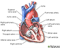
Heart - section through the middle - illustration
The interior of the heart is composed of valves, chambers, and associated vessels.
Heart - section through the middle
illustration
-
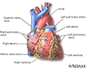
Heart - front view - illustration
The external structures of the heart include the ventricles, atria, arteries and veins. Arteries carry blood away from the heart while veins carry blood into the heart. The vessels colored blue indicate the transport of blood with relatively low content of oxygen and high content of carbon dioxide. The vessels colored red indicate the transport of blood with relatively high content of oxygen and low content of carbon dioxide.
Heart - front view
illustration
-
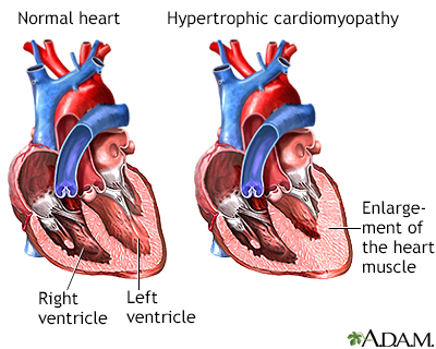
Hypertrophic cardiomyopathy - illustration
Hypertrophic cardiomyopathy is the thickening of the muscles that make up the heart. The thickening may interfere with the normal functioning of the heart by narrowing the outflow of the ventricle; reducing the ability of the heart to relax and fill with blood during the relaxation phase; or reducing the ability of the valves of the heart to function properly. Any situation that increases the contraction or rate of contraction of the heart muscle can worsen these symptoms.
Hypertrophic cardiomyopathy
illustration


-
Heart - section through the middle - illustration
The interior of the heart is composed of valves, chambers, and associated vessels.
Heart - section through the middle
illustration
-
Heart - front view - illustration
The external structures of the heart include the ventricles, atria, arteries and veins. Arteries carry blood away from the heart while veins carry blood into the heart. The vessels colored blue indicate the transport of blood with relatively low content of oxygen and high content of carbon dioxide. The vessels colored red indicate the transport of blood with relatively high content of oxygen and low content of carbon dioxide.
Heart - front view
illustration
-
Hypertrophic cardiomyopathy - illustration
Hypertrophic cardiomyopathy is the thickening of the muscles that make up the heart. The thickening may interfere with the normal functioning of the heart by narrowing the outflow of the ventricle; reducing the ability of the heart to relax and fill with blood during the relaxation phase; or reducing the ability of the valves of the heart to function properly. Any situation that increases the contraction or rate of contraction of the heart muscle can worsen these symptoms.
Hypertrophic cardiomyopathy
illustration
Review Date: 5/8/2022
Reviewed By: Michael A. Chen, MD, PhD, Associate Professor of Medicine, Division of Cardiology, Harborview Medical Center, University of Washington Medical School, Seattle, WA. Also reviewed by David C. Dugdale, MD, Medical Director, Brenda Conaway, Editorial Director, and the A.D.A.M. Editorial team.
 All rights reserved.