
Silicosis
Acute silicosis; Chronic silicosis; Accelerated silicosis; Progressive massive fibrosis; Conglomerate silicosis; SilicoproteinosisSilicosis is a lung disease caused by breathing in (inhaling) silica dust.
Causes
Silica is a common, naturally-occurring crystal. It is found in most rock beds. Silica dust forms during mining, quarrying, tunneling, and working with certain metal ores. Silica is a main part of sand, so glass workers and sand-blasters are also exposed to silica.
Three types of silicosis occur:
- Chronic silicosis results from long-term exposure (more than 20 years) to low amounts of silica dust. The silica dust causes swelling in the lungs and chest lymph nodes. This disease may cause people to have trouble breathing. This is the most common form of silicosis.
- Accelerated silicosis occurs after exposure to larger amounts of silica over a shorter period of time (3 to 10 years). Swelling in the lungs and symptoms occur faster than in simple silicosis.
- Acute silicosis results from short-term exposure to very large amounts of silica. The lungs become very inflamed and can fill with fluid, causing severe shortness of breath and a low blood oxygen level.
People who work in jobs where they are exposed to silica dust are at risk. These jobs include:
- Abrasives manufacturing
- Glass manufacturing
- Mining
- Quarrying
- Road and building construction
- Sand blasting
- Stone cutting
Intense exposure to silica can cause disease within a year. But it usually takes at least 10 years of exposure before symptoms occur. Silicosis has become less common since the Occupational Safety and Health Administration (OSHA) created regulations requiring the use of protective equipment, which limits the amount of silica dust workers inhale.
Symptoms
Symptoms include:
-
Cough
Cough
Coughing is an important way to keep your throat and airways clear. But too much coughing may mean you have a disease or disorder. Some coughs are d...
 ImageRead Article Now Book Mark Article
 - Shortness of breath
- Weight loss
Exams and Tests
Your health care provider will take a medical history. You'll be asked about your jobs (past and present), hobbies, and other activities that may have exposed you to silica. Your provider will also do a physical exam.
Tests to confirm the diagnosis and rule out similar diseases include:
-
Chest x-ray
Chest x-ray
A chest x-ray is an x-ray of the chest, lungs, heart, large arteries, ribs, and diaphragm.
 ImageRead Article Now Book Mark Article
 -
Chest CT scan
Chest CT scan
A chest CT (computed tomography) scan is an imaging method that uses x-rays to create cross-sectional pictures of the chest and upper abdomen....
 ImageRead Article Now Book Mark Article
 -
Lung function tests
Lung function tests
Pulmonary function tests are a group of tests that measure breathing and how well the lungs are functioning.
 ImageRead Article Now Book Mark Article
 -
Tuberculosis (TB) tests
Tuberculosis (TB) tests
The PPD skin test is a method used to diagnose silent (latent) tuberculosis (TB) infection. PPD stands for purified protein derivative.
 ImageRead Article Now Book Mark Article
 - Blood tests for connective tissue diseases
Treatment
There is no specific treatment for silicosis. Removing the source of silica exposure is important to prevent the disease from getting worse. Supportive treatment includes cough medicine, bronchodilators, and oxygen if needed. Antibiotics are prescribed for respiratory infections as needed.
Treatment also includes limiting exposure to irritants and quitting smoking.
People with silicosis are at high risk for developing TB. Silica is believed to interfere with the body's immune response to the bacteria that cause TB. Skin tests to check for exposure to TB should be done regularly. Those with a positive skin test should be treated with anti-TB medicines. Any change in the appearance of the chest x-ray may be a sign of TB.
People with severe silicosis may need to have a lung transplant in rare cases.
Support Groups
Joining a support group where you can meet other people with silicosis or related diseases can help you understand your disease and adapt to its treatments.
Outlook (Prognosis)
Outcome varies, depending on the amount of damage to the lungs.
Possible Complications
Silicosis can lead to the following health problems:
- Connective tissue disease, including rheumatoid arthritis, scleroderma (also called progressive systemic sclerosis), and systemic lupus erythematosus
Rheumatoid arthritis
Rheumatoid arthritis (RA) is a disease that leads to inflammation of the joints and surrounding tissues. It is a long-term disease. It can also aff...
 ImageRead Article Now Book Mark Article
Scleroderma
Scleroderma is a disease that involves the buildup of fibrous tissue in the skin and elsewhere in the body. It also damages the cells that line the ...
 ImageRead Article Now Book Mark Article
Systemic lupus erythematosus
Systemic lupus erythematosus (SLE) is an autoimmune disease. In this disease, the immune system of the body mistakenly attacks healthy tissue. It c...
 ImageRead Article Now Book Mark Article
 - Lung cancer
- Progressive massive fibrosis
-
Respiratory failure
Respiratory failure
Respiratory acidosis is a condition that occurs when your lungs can't remove all of the carbon dioxide produced by your body. This causes the blood ...
 ImageRead Article Now Book Mark Article
 - Tuberculosis
When to Contact a Medical Professional
Contact your provider if you suspect that you have been exposed to silica at work and you have breathing problems. Having silicosis makes it easier for you to develop lung infections. Talk to your provider about getting the flu, pneumonia, and other recommended vaccines.
If you've been diagnosed with silicosis, call your provider right away if you develop a cough, shortness of breath, fever, or other signs of a lung infection, especially if you think you have the flu. Since your lungs are already damaged, it's very important to have the infection treated promptly. This will prevent breathing problems from becoming severe, as well as further damage to your lungs.
Prevention
If you work in a high-risk occupation or have a high-risk hobby, always wear a dust mask and do not smoke. You might also want to use other protection recommended by OSHA, such as a respirator.
References
Go LHT, Cohen RA. Pneumoconioses. In: Broaddus VC, Ernst JD, King TE, et al, eds. Murray and Nadel's Textbook of Respiratory Medicine. 7th ed. Philadelphia, PA: Elsevier; 2022:chap 101.
Tarlo SM. Occupational lung disease. In: Goldman L, Schafer AI, eds. Goldman-Cecil Medicine. 26th ed. Philadelphia, PA: Elsevier; 2020:chap 87.
-
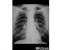
Coal worker's lungs - chest x-ray - illustration
This chest x-ray shows coal worker's lungs. There are diffuse, small, light areas on both sides (1 to 3 mm) in all parts of the lungs. Diseases that may result in an x-ray like this include simple coal workers pneumoconiosis (CWP) - stage I, simple silicosis, miliary tuberculosis, histiocytosis X (eosinophilic granuloma), and other diffuse infiltrate pulmonary diseases.
Coal worker's lungs - chest x-ray
illustration
-
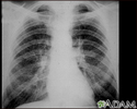
Coal workers pneumoconiosis - stage II - illustration
This chest x-ray shows stage II coal workers pneumoconiosis (CWP). There are diffuse, small light areas on both sides of the lungs. Other diseases that may explain these x-ray findings include simple silicosis, disseminated tuberculosis, metastatic lung cancer, and other diffuse, infiltrative pulmonary diseases.
Coal workers pneumoconiosis - stage II
illustration
-
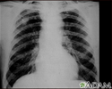
Coal workers pneumoconiosis - stage II - illustration
This chest x-ray shows coal workers pneumoconiosis - stage II. There are diffuse, small (2 to 4 mm each), light areas throughout both lungs. In the right upper lung (seen on the left side of the picture), there is a light area (measuring approximately 2 cm by 4 cm) with poorly defined borders, representing coalescence (merging together) of previously distinct light areas. Diseases which may explain these x-ray findings include simple coal workers pneumoconiosis (CWP) - stage II, silico-tuberculosis, disseminated tuberculosis, metastatic lung cancer, and other diffuse infiltrative pulmonary diseases.
Coal workers pneumoconiosis - stage II
illustration
-
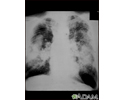
Coal workers pneumoconiosis, complicated - illustration
This picture shows complicated coal workers pneumoconiosis. There are diffuse, massive light areas that run together in the upper and middle parts of both lungs. These are superimposed on a background of small and poorly distinguishable light areas that are diffuse and located in both lungs. Diseases which may explain these x-ray findings include, but are not limited to complicated coal workers pneumoconiosis (CWP), silico-tuberculosis, and metastatic lung cancer.
Coal workers pneumoconiosis, complicated
illustration
-
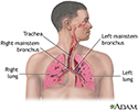
Respiratory system - illustration
Air is breathed in through the nasal passageways, travels through the trachea and bronchi to the lungs.
Respiratory system
illustration
-
Coal worker's lungs - chest x-ray - illustration
This chest x-ray shows coal worker's lungs. There are diffuse, small, light areas on both sides (1 to 3 mm) in all parts of the lungs. Diseases that may result in an x-ray like this include simple coal workers pneumoconiosis (CWP) - stage I, simple silicosis, miliary tuberculosis, histiocytosis X (eosinophilic granuloma), and other diffuse infiltrate pulmonary diseases.
Coal worker's lungs - chest x-ray
illustration
-
Coal workers pneumoconiosis - stage II - illustration
This chest x-ray shows stage II coal workers pneumoconiosis (CWP). There are diffuse, small light areas on both sides of the lungs. Other diseases that may explain these x-ray findings include simple silicosis, disseminated tuberculosis, metastatic lung cancer, and other diffuse, infiltrative pulmonary diseases.
Coal workers pneumoconiosis - stage II
illustration
-
Coal workers pneumoconiosis - stage II - illustration
This chest x-ray shows coal workers pneumoconiosis - stage II. There are diffuse, small (2 to 4 mm each), light areas throughout both lungs. In the right upper lung (seen on the left side of the picture), there is a light area (measuring approximately 2 cm by 4 cm) with poorly defined borders, representing coalescence (merging together) of previously distinct light areas. Diseases which may explain these x-ray findings include simple coal workers pneumoconiosis (CWP) - stage II, silico-tuberculosis, disseminated tuberculosis, metastatic lung cancer, and other diffuse infiltrative pulmonary diseases.
Coal workers pneumoconiosis - stage II
illustration
-
Coal workers pneumoconiosis, complicated - illustration
This picture shows complicated coal workers pneumoconiosis. There are diffuse, massive light areas that run together in the upper and middle parts of both lungs. These are superimposed on a background of small and poorly distinguishable light areas that are diffuse and located in both lungs. Diseases which may explain these x-ray findings include, but are not limited to complicated coal workers pneumoconiosis (CWP), silico-tuberculosis, and metastatic lung cancer.
Coal workers pneumoconiosis, complicated
illustration
-
Respiratory system - illustration
Air is breathed in through the nasal passageways, travels through the trachea and bronchi to the lungs.
Respiratory system
illustration
Review Date: 5/3/2023
Reviewed By: Denis Hadjiliadis, MD, MHS, Paul F. Harron, Jr. Professor of Medicine, Pulmonary, Allergy, and Critical Care, Perelman School of Medicine, University of Pennsylvania, Philadelphia, PA. Also reviewed by David C. Dugdale, MD, Medical Director, Brenda Conaway, Editorial Director, and the A.D.A.M. Editorial team.
 All rights reserved.