
Pulmonary aspergilloma
Fungus ball; Mycetoma; Aspergilloma; Aspergillosis - pulmonary aspergillomaPulmonary aspergilloma is a mass caused by an infection by the fungus aspergillus. It usually grows in preexisting lung cavities. The infection can also appear in the brain, kidney, or other organs.
Causes
Aspergillosis is an infection caused by the fungus aspergillus. Aspergillomas are formed when the fungus grows in a clump in a lung cavity. The cavity is often created by a previous condition. Cavities in the lung may be caused by diseases such as:
Aspergillosis
Aspergillosis is an infection or allergic response due to the aspergillus fungus.

- Tuberculosis
Tuberculosis
Pulmonary tuberculosis (TB) is a contagious bacterial infection that involves the lungs. It may spread to other organs.
 ImageRead Article Now Book Mark Article
 - Coccidioidomycosis
Coccidioidomycosis
Valley fever is an infection that occurs when the spores of the fungus Coccidioides immitis or Coccidioides posadasii enter your body through the lun...
 ImageRead Article Now Book Mark Article
 - Cystic fibrosis
Cystic fibrosis
Cystic fibrosis is a disease that causes thick, sticky mucus to build up in the lungs, digestive tract, and other areas of the body. It is one of th...
 ImageRead Article Now Book Mark Article
 - Histoplasmosis
Histoplasmosis
Histoplasmosis is an infection that occurs from breathing in the spores of the fungus Histoplasma capsulatum.
 ImageRead Article Now Book Mark Article
 -
Lung abscess
Abscess
An abscess is a collection of pus in any part of the body. In most cases, the area around an abscess is swollen and inflamed.
 ImageRead Article Now Book Mark Article
 - Lung cancer
Lung cancer
Lung cancer is cancer that starts in the lungs. The lungs are located in the chest. When you breathe, air goes through your nose, down your windpipe...
ImageRead Article Now Book Mark Article - Sarcoidosis
Sarcoidosis
Sarcoidosis is a disease in which inflammation occurs in the lymph nodes, lungs, liver, eyes, skin, and/or other tissues.
 ImageRead Article Now Book Mark Article
The most common species of fungus that causes disease in humans is Aspergillus fumigatus.
Aspergillus is a common fungus. It grows on dead leaves, stored grain, bird droppings, compost piles, and other decaying vegetation.
Symptoms
You may not have any symptoms. When symptoms do develop, they can include:
- Chest pain
Chest pain
Chest pain is discomfort or pain that you feel anywhere along the front of your body between your neck and upper abdomen.
 ImageRead Article Now Book Mark Article
 - Cough
- Coughing up blood, which can be a life-threatening sign
Coughing up blood
Coughing up blood is the spitting up of blood or bloody mucus from the lungs and throat (respiratory tract). Hemoptysis is the medical term for cough...
 ImageRead Article Now Book Mark Article
 - Fatigue
- Fever
- Unintentional weight loss
Unintentional weight loss
Unexplained weight loss is a decrease in body weight, when you did not try to lose the weight on your own. Many people gain and lose weight. Uninten...
Read Article Now Book Mark Article
Exams and Tests
Your health care provider may suspect you have a fungal infection after x-rays of your lungs show the ball of fungus. Other tests that may be done include:
x-rays
X-rays are a type of electromagnetic radiation, just like visible light. An x-ray machine sends individual x-ray waves through the body. The images...

- Biopsy of lung tissue
Biopsy
A biopsy is the removal of a small piece of tissue for lab examination.
 ImageRead Article Now Book Mark Article
 - Blood test for presence of aspergillus in the body (galactomannan)
- Blood test to detect immune response to aspergillus (aspergillus precipitin)
Aspergillus precipitin
Aspergillosis precipitin is a laboratory test to detect antibodies in the blood resulting from exposure to the fungus aspergillus.
 ImageRead Article Now Book Mark Article
 - Bronchoscopy or bronchoscopy with lavage
Bronchoscopy
Bronchoscopy is a test to view the airways and diagnose lung disease. It may also be used during the treatment of some lung conditions.
 ImageRead Article Now Book Mark Article
 - Chest CT
Chest CT
A chest CT (computed tomography) scan is an imaging method that uses x-rays to create cross-sectional pictures of the chest and upper abdomen....
 ImageRead Article Now Book Mark Article
 - Sputum culture
Sputum culture
Routine sputum culture is a laboratory test that looks for germs that cause infection. Sputum is the material that comes up from air passages when y...
 ImageRead Article Now Book Mark Article
Treatment
Many people never develop symptoms. Often, no treatment is needed, unless you are coughing up blood.
Sometimes, antifungal medicines may be used.
If you have bleeding in the lungs, your provider may recommend a test to inject dye into the blood vessels (angiography) to find the site of bleeding. The bleeding is stopped by either:
Angiography
CT angiography combines a CT scan with the injection of dye. This technique is able to create pictures of the blood vessels in the chest and upper a...

- Surgery to remove the aspergilloma
- A procedure that inserts material into the blood vessels to stop the bleeding (embolization)
Embolization
Endovascular embolization is a procedure to treat abnormal blood vessels in the brain and other parts of the body. It is an alternative to open surg...
Read Article Now Book Mark Article
Outlook (Prognosis)
The outcome can be good for many people. However, it depends on the severity of the condition and your overall health.
Surgery may be very successful in some cases, but it is complex and can have a high risk of serious complications.
Possible Complications
Complications of pulmonary aspergilloma may include:
- Difficulty breathing that gets worse
Difficulty breathing
Breathing difficulty may involve:Difficult breathing Uncomfortable breathingFeeling like you are not getting enough air
ImageRead Article Now Book Mark Article - Massive bleeding from the lung
- Spread of the infection
When to Contact a Medical Professional
Contact your provider if you cough up blood, and be sure to mention any other symptoms that have developed.
Prevention
People who have had related lung infections or who have weakened immune systems should try to avoid environments where the aspergillus fungus is found.
References
Patterson TF, Thompson GR 3rd, Denning DW, et al. Practice guidelines for the diagnosis and management of aspergillosis: 2016 update by the Infectious Diseases Society of America. Clin Infect Dis. 2016;63(4):e1-e60. PMID: 27365388 pubmed.ncbi.nlm.nih.gov/27365388/.
Saullo JL, Alexander BD. Fungal infections: opportunistic. In: Broaddus VC, Ernst JD, King TE, et al, eds. Murray and Nadel's Textbook of Respiratory Medicine. 7th ed. Philadelphia, PA: Elsevier; 2022:chap 57.
Walsh TJ, Patterson TF. Aspergillosis. In: Goldman L, Cooney KA, eds. Goldman-Cecil Medicine. 27th ed. Philadelphia, PA: Elsevier; 2024:chap 311.
Lungs - illustration
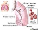
The major features of the lungs include the bronchi, the bronchioles and the alveoli. The alveoli are the microscopic blood vessel-lined sacks in which oxygen and carbon dioxide gas are exchanged.
Lungs
illustration
Pulmonary nodule - front view chest x-ray - illustration
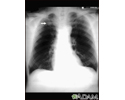
This x-ray shows a single lesion (pulmonary nodule) in the upper right lung (seen as a light area on the left side of the picture). The nodule has distinct borders (well-defined) and is uniform in density. Tuberculosis (TB) and other diseases can cause this type of lesion.
Pulmonary nodule - front view chest x-ray
illustration
Pulmonary nodule, solitary - CT scan - illustration
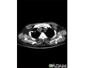
This CT scan shows a single lesion (pulmonary nodule) in the right lung. This nodule is seen as the light circle in the upper portion of the dark area on the left side of the picture. A normal lung would look completely black in a CT scan.
Pulmonary nodule, solitary - CT scan
illustration
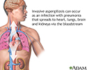
Aspergilloma - illustration
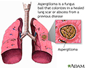
Aspergillosis is an acute pulmonary infection caused by the aspergillus fungus. Aspergillus can cause illness three ways an allergic reaction in asthmatics, a colonization in scarred lung tissue, and an invasive infection with pneumonia, which can affect the heart, lungs, brain and kidneys.
Aspergilloma
illustration
Pulmonary aspergillosis - illustration
Aspergillosis is an acute pulmonary infection caused by the aspergillus fungus. Aspergillus can cause illness three ways an allergic reaction in asthmatics; a colonization in scarred lung tissue; and an invasive infection with pneumonia which can affect the heart, lungs, brain and kidneys.
Pulmonary aspergillosis
illustration
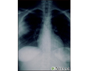
Aspergillosis - chest X-ray - illustration
Aspergillosis is a fungal infection. The fungus invades and destroys tissue. This type of infection usually occurs in immunocompromised individuals. Here, a chest x-ray shows that the fungus has invaded the lung tissue. The lungs are usually seen as black areas on an x-ray. The cloudiness on the left side of this x-ray is caused by the fungus.
Aspergillosis - chest X-ray
illustration
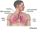
Respiratory system - illustration
Air is breathed in through the nasal passageways, travels through the trachea and bronchi to the lungs.
Respiratory system
illustration
Lungs - illustration
The major features of the lungs include the bronchi, the bronchioles and the alveoli. The alveoli are the microscopic blood vessel-lined sacks in which oxygen and carbon dioxide gas are exchanged.
Lungs
illustration
Pulmonary nodule - front view chest x-ray - illustration
This x-ray shows a single lesion (pulmonary nodule) in the upper right lung (seen as a light area on the left side of the picture). The nodule has distinct borders (well-defined) and is uniform in density. Tuberculosis (TB) and other diseases can cause this type of lesion.
Pulmonary nodule - front view chest x-ray
illustration
Pulmonary nodule, solitary - CT scan - illustration
This CT scan shows a single lesion (pulmonary nodule) in the right lung. This nodule is seen as the light circle in the upper portion of the dark area on the left side of the picture. A normal lung would look completely black in a CT scan.
Pulmonary nodule, solitary - CT scan
illustration
Aspergilloma - illustration
Aspergillosis is an acute pulmonary infection caused by the aspergillus fungus. Aspergillus can cause illness three ways an allergic reaction in asthmatics, a colonization in scarred lung tissue, and an invasive infection with pneumonia, which can affect the heart, lungs, brain and kidneys.
Aspergilloma
illustration
Pulmonary aspergillosis - illustration
Aspergillosis is an acute pulmonary infection caused by the aspergillus fungus. Aspergillus can cause illness three ways an allergic reaction in asthmatics; a colonization in scarred lung tissue; and an invasive infection with pneumonia which can affect the heart, lungs, brain and kidneys.
Pulmonary aspergillosis
illustration
Aspergillosis - chest X-ray - illustration
Aspergillosis is a fungal infection. The fungus invades and destroys tissue. This type of infection usually occurs in immunocompromised individuals. Here, a chest x-ray shows that the fungus has invaded the lung tissue. The lungs are usually seen as black areas on an x-ray. The cloudiness on the left side of this x-ray is caused by the fungus.
Aspergillosis - chest X-ray
illustration
Respiratory system - illustration
Air is breathed in through the nasal passageways, travels through the trachea and bronchi to the lungs.
Respiratory system
illustration
Review Date: 8/29/2024
Reviewed By: Jatin M. Vyas, MD, PhD, Professor in Medicine, Harvard Medical School; Associate in Medicine, Division of Infectious Disease, Department of Medicine, Massachusetts General Hospital, Boston, MA. Also reviewed by David C. Dugdale, MD, Medical Director, Brenda Conaway, Editorial Director, and the A.D.A.M. Editorial team.
 All rights reserved.