
Hemothorax
Hemothorax is a collection of blood in the space between the chest wall and the lung (the pleural cavity).
Causes
The most common cause of hemothorax is chest trauma. It can sometimes be accompanied by air in the pleural cavity that may let the lung partially collapse (pneumothorax). Hemothorax can also occur in people who have:
- A blood clotting defect
- Chest (thoracic) or heart surgery
- Death of lung tissue (pulmonary infarction)
- Lung or pleural cancer -- primary or secondary (metastatic, or from another site)
Cancer
Cancer is the uncontrolled growth of abnormal cells in the body. Cancerous cells are also called malignant cells.
Read Article Now Book Mark Article - A tear in a blood vessel when placing a central venous catheter or when associated with severe high blood pressure
- Tuberculosis
Symptoms
Symptoms include:
- Shortness of breath
Shortness of breath
Breathing difficulty may involve:Difficult breathing Uncomfortable breathingFeeling like you are not getting enough air
 ImageRead Article Now Book Mark Article
 - Rapid, shallow breathing
- Chest pain
Chest pain
Chest pain is discomfort or pain that you feel anywhere along the front of your body between your neck and upper abdomen.
 ImageRead Article Now Book Mark Article
 - Low blood pressure (shock)
Low blood pressure
Low blood pressure occurs when blood pressure is below normal. This means the heart, brain, and other parts of the body may not get enough blood. I...
 ImageRead Article Now Book Mark Article
 - Pale, cool and clammy skin
- Rapid heart rate
Rapid heart rate
A bounding pulse is a strong throbbing felt over one of the arteries in the body. It is due to a forceful heartbeat.
 ImageRead Article Now Book Mark Article
 - Restlessness
Restlessness
Agitation is an unpleasant state of extreme arousal. An agitated person may feel stirred up, excited, tense, confused, or irritable.
Read Article Now Book Mark Article - Anxiety
Anxiety
Stress is a feeling of emotional or physical tension. It can come from any event or thought that makes you feel frustrated, angry, or nervous. Stres...
 ImageRead Article Now Book Mark Article
Exams and Tests
Your health care provider may note decreased or absent breath sounds on the affected side. Signs or findings of hemothorax may be seen on the following tests:
- Chest x-ray
Chest x-ray
A chest x-ray is an x-ray of the chest, lungs, heart, large arteries, ribs, and diaphragm.
 ImageRead Article Now Book Mark Article
 - CT scan
CT scan
A computed tomography (CT) scan is an imaging method that uses x-rays to create pictures of cross-sections of the body. Related tests include:Abdomin...
 ImageRead Article Now Book Mark Article
 - Thoracentesis (drainage of pleural fluid through a needle or catheter)
Thoracentesis
Thoracentesis is a procedure to remove fluid from the space between the lining of the outside of the lungs (pleura) and the wall of the chest....
Read Article Now Book Mark Article - Thoracostomy (drainage of pleural fluid through a chest tube)
Treatment
The goal of treatment is to get the person stable, stop the bleeding, and remove the blood and air in the pleural space.
- A chest tube is inserted through the chest wall between the ribs to drain the blood and air.
- It is left in place and attached to suction for several days to re-expand the lung.
If a chest tube alone does not control the bleeding, surgery (thoracotomy) may be needed to stop the bleeding.
Thoracotomy
Lung surgery is surgery done to repair or remove lung tissue. There are many common lung surgeries, including:Biopsy of an unknown growth in or arou...

The cause of the hemothorax will be also treated. The underlying lung may have collapsed. This can lead to breathing difficulty. In people who have had an injury, chest tube drainage may be all that is needed. Surgery may not be necessary.
WHAT TO EXPECT AT THE EMERGENCY DEPARTMENT
The provider will measure and monitor the person's vital signs, including oxygen saturation, pulse, breathing rate, and blood pressure. Symptoms will be treated as needed. The person may receive:
- Breathing support -- This may include oxygen, non-invasive airway pressure support such as BIPAP, or endotracheal intubation (placement of a breathing tube through the mouth or nose into the airway) and placement on a ventilator (life support breathing machine)
- Blood tests and possible blood transfusion
- Chest tube (tube through the skin and muscles between the ribs into the space around the lungs) if there is lung collapse
- CT scan
- Analysis of the pleural fluid
- Electrocardiogram (ECG)
- Fluids given through the vein (IV)
- Medicines to treat symptoms
- X-rays of chest and abdomen or other parts of the body if there are additional injuries
Outlook (Prognosis)
The outcome depends on the cause of the hemothorax, the amount of blood loss and how quickly treatment is given.
In the case of major trauma, the outcome will additionally depend on the severity of the injury and the rate of bleeding.
Possible Complications
Complications may include:
- Collapsed lung, or pneumothorax, leading to respiratory failure (inability to breathe properly)
Collapsed lung
A collapsed lung occurs when air escapes from the lung. The air then fills the space outside of the lung between the lung and chest wall. This buil...
ImageRead Article Now Book Mark ArticlePneumothorax
A collapsed lung occurs when air escapes from the lung. The air then fills the space outside of the lung between the lung and chest wall. This buil...
ImageRead Article Now Book Mark ArticleRespiratory
The words "respiratory" and "respiration" refer to the lungs and breathing.
ImageRead Article Now Book Mark Article - Fibrosis or scarring of the pleural membranes and underlying lung tissue
- Infection of the pleural fluid (empyema)
Empyema
Empyema is a collection of pus in the space between the lung and the inner surface of the chest wall (pleural space).
ImageRead Article Now Book Mark Article - Shock and death in severe circumstances
Shock
Shock is a life-threatening condition that occurs when the body is not getting enough blood flow. Lack of blood flow means the cells and organs do n...
 ImageRead Article Now Book Mark Article
When to Contact a Medical Professional
Call 911 or the local emergency number if you have:
- Any potentially serious injury to the chest
- Chest pain
- Severe jaw, neck, shoulder or arm pain
- Difficulty breathing or shortness of breath
Go to the emergency room or call the local emergency number (such as 911) if you have:
- Dizziness, lightheadedness, fever and cough, or a feeling of heaviness in your chest
Prevention
Use safety measures (such as seat belts) to avoid injury. Depending on the cause, a hemothorax may not be preventable.
References
Cameron J. Trauma and emergency care. In: Cameron J, ed. Current Surgical Therapy. 14th ed. Philadelphia, PA: Elsevier; 2023:chap 17.
Friedberg J, Stewart SJ. Hemothorax. In: Broaddus VC, Ernst JD, King TE, et al, eds. Murray and Nadel's Textbook of Respiratory Medicine. 7th ed. Philadelphia, PA: Elsevier; 2022:chap 113.
Raja AS. Thoracic trauma. In: Walls RM, ed. Rosen's Emergency Medicine: Concepts and Clinical Practice. 10th ed. Philadelphia, PA: Elsevier; 2023:chap 37.
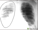
Aortic rupture - chest X-ray - illustration
Aortic rupture (a tear in the aorta, which is the major artery coming from the heart) can be seen on a chest X-ray. In this case, it was caused by a traumatic perforation of the thoracic aorta. This is how the X-ray appears when the chest is full of blood (right-sided hemothorax) seen here as cloudiness on the left side of the picture.
Aortic rupture - chest X-ray
illustration
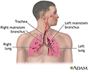
Respiratory system - illustration
Air is breathed in through the nasal passageways, travels through the trachea and bronchi to the lungs.
Respiratory system
illustration
Chest tube insertion - series - Pleural cavity
Presentation
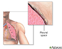
 and the chest cavity. </p>")
, bleeding into the chest (hemothorax), after surgery or trauma in the chest (pneumothorax or hemothorax), lung abscesses or pus in the chest (empyema). </p>")
. The patient may also be sedated. The chest tube is inserted between the ribs into the chest and is connected to a bottle or canister that contains sterile water. Suction is attached to the system to encourage drainage. A stitch (suture) and adhesive tape is used to keep the tube in place. The chest tube usually remains in place until the X-rays show that all the blood, fluid, or air has drained from the chest and the lung has fully re-expanded. When the chest tube is no longer needed, it can be easily removed, usually without the need for medications to sedate or numb the patient. Medications may be used to prevent or treat infection (antibiotics).</p>")

Aortic rupture - chest X-ray - illustration
Aortic rupture (a tear in the aorta, which is the major artery coming from the heart) can be seen on a chest X-ray. In this case, it was caused by a traumatic perforation of the thoracic aorta. This is how the X-ray appears when the chest is full of blood (right-sided hemothorax) seen here as cloudiness on the left side of the picture.
Aortic rupture - chest X-ray
illustration
Respiratory system - illustration
Air is breathed in through the nasal passageways, travels through the trachea and bronchi to the lungs.
Respiratory system
illustration
Chest tube insertion - series - Pleural cavity
Presentation
Review Date: 7/12/2024
Reviewed By: Jesse Borke, MD, CPE, FAAEM, FACEP, Attending Physician at Kaiser Permanente, Orange County, CA. Also reviewed by David C. Dugdale, MD, Medical Director, Brenda Conaway, Editorial Director, and the A.D.A.M. Editorial team.
 All rights reserved.