
Collapsed lung (pneumothorax)
Air around the lung; Air outside the lung; Pneumothorax dropped lung; Spontaneous pneumothoraxA collapsed lung occurs when air escapes from the lung. The air then fills the space outside of the lung between the lung and chest wall. This buildup of air puts pressure on the lung, so it cannot expand as much as it normally does when you take a breath.
The medical name of this condition is pneumothorax.
Causes
Collapsed lung can be caused by an injury to the lung. Injuries can include a gunshot or knife wound to the chest, rib fracture, or certain medical procedures.
In some cases, a collapsed lung is caused by air blisters of the lung (blebs) that break open, sending air into the space around the lung. This can result from air pressure changes such as when scuba diving or traveling to a high altitude.
Tall, thin people and smokers are more at risk for a collapsed lung.
Lung diseases can also increase the chance of getting a collapsed lung. These include:
- Asthma
Asthma
Asthma is a chronic disease that causes the airways of the lungs to swell and narrow. It leads to breathing difficulty such as wheezing, shortness o...
 ImageRead Article Now Book Mark Article
 - Chronic obstructive pulmonary disease (COPD)
Chronic obstructive pulmonary disease (...
Chronic obstructive pulmonary disease (COPD) is a common lung disease. Having COPD makes it hard to breathe. There are two main forms of COPD:Chroni...
 ImageRead Article Now Book Mark Article
 - Cystic fibrosis
Cystic fibrosis
Cystic fibrosis is a disease that causes thick, sticky mucus to build up in the lungs, digestive tract, and other areas of the body. It is one of th...
 ImageRead Article Now Book Mark Article
 - Tuberculosis
Tuberculosis
Pulmonary tuberculosis (TB) is a contagious bacterial infection that involves the lungs. It may spread to other organs.
 ImageRead Article Now Book Mark Article
 - Whooping cough
Whooping cough
Pertussis is a highly contagious bacterial disease that causes uncontrollable, violent coughing. The coughing can make it hard to breathe. A deep "...
 ImageRead Article Now Book Mark Article
In some cases, a collapsed lung occurs without any cause. This is called a spontaneous collapsed lung or spontaneous pneumothorax.
Symptoms
Common symptoms of a collapsed lung include:
- Sharp chest or shoulder pain, made worse by a deep breath or a cough
- Shortness of breath
Shortness of breath
Breathing difficulty may involve:Difficult breathing Uncomfortable breathingFeeling like you are not getting enough air
 ImageRead Article Now Book Mark Article
 - Nasal flaring (from shortness of breath)
Nasal flaring
Nasal flaring occurs when the nostrils widen while breathing. It is often a sign of trouble breathing.
 ImageRead Article Now Book Mark Article
A larger pneumothorax causes more severe symptoms, including:
- Bluish color of the skin due to lack of oxygen
Bluish color
A bluish color to the skin or mucous membrane is usually due to a lack of oxygen in the blood. The medical term is cyanosis.
 ImageRead Article Now Book Mark Article
 - Chest tightness
- Lightheadedness and near fainting
- Easy fatigue
Easy fatigue
Fatigue is a feeling of weariness, tiredness, or lack of energy.
Read Article Now Book Mark Article - Abnormal breathing patterns or increased effort of breathing
- Rapid heart rate
Rapid heart rate
A bounding pulse is a strong throbbing felt over one of the arteries in the body. It is due to a forceful heartbeat.
 ImageRead Article Now Book Mark Article
 - Shock and collapse
Shock
Shock is a life-threatening condition that occurs when the body is not getting enough blood flow. Lack of blood flow means the cells and organs do n...
 ImageRead Article Now Book Mark Article
Exams and Tests
The health care provider will listen to your breathing with a stethoscope. If you have a collapsed lung, there are decreased breath sounds or no breath sounds on the affected side. You may also have low blood pressure.
Tests that may be ordered include:
- Chest x-ray
Chest x-ray
A chest x-ray is an x-ray of the chest, lungs, heart, large arteries, ribs, and diaphragm.
 ImageRead Article Now Book Mark Article
 - Arterial blood gases and other blood tests
Arterial blood gases
Blood gases are a measurement of how much oxygen and carbon dioxide are in your blood. They also determine the acidity (pH) of your blood.
 ImageRead Article Now Book Mark Article
 - CT scan if other injuries or conditions are suspected
CT scan
A chest CT (computed tomography) scan is an imaging method that uses x-rays to create cross-sectional pictures of the chest and upper abdomen....
 ImageRead Article Now Book Mark Article
 - Electrocardiogram (ECG)
Electrocardiogram
An electrocardiogram (ECG) is a test that records the electrical activity of the heart.
 ImageRead Article Now Book Mark Article
Treatment
A small pneumothorax may go away on its own over time. You may only need oxygen treatment and rest.
The provider may use a needle to allow the air to escape from around the lung so it can expand more fully. You may be allowed to go home if you live near the hospital.
If you have a large pneumothorax, a chest tube will be placed between the ribs into the space around the lungs to help drain the air and allow the lung to re-expand. The chest tube may be left in place for several days and you may need to stay in the hospital. If a small chest tube or flutter valve is used, you may be able to go home. You will need to return to the hospital to have the tube or valve removed.
Chest tube
A chest tube is a hollow, flexible tube placed into the chest. It acts as a drain. Chest tubes drain blood, fluid, or air from around your lungs, he...

Some people with a collapsed lung need extra oxygen.
Lung surgery may be needed to treat collapsed lung or to prevent future episodes. The area where the leak occurred may be repaired. Sometimes, a special chemical is placed into the area of the collapsed lung. This chemical causes a scar to form. This procedure is called pleurodesis.
Lung surgery
Lung surgery is surgery done to repair or remove lung tissue. There are many common lung surgeries, including:Biopsy of an unknown growth in or arou...

Outlook (Prognosis)
If you have a spontaneous collapsed lung, you are more likely to have another one in the future if you:
- Are tall and thin
- Continue to smoke
- Have had two collapsed lung episodes in the past
How well you do after having a collapsed lung depends on what caused it.
Possible Complications
Complications may include any of the following:
- Another collapsed lung in the future
- Shock, if there are serious injuries or infection, severe inflammation, or fluid in the lung develops
Shock
Shock is a life-threatening condition that occurs when the body is not getting enough blood flow. Lack of blood flow means the cells and organs do n...
ImageRead Article Now Book Mark Article
When to Contact a Medical Professional
Contact your provider if you have symptoms of a collapsed lung, especially if you have had one before.
Prevention
There is no known way to prevent a collapsed lung. Following standard procedure can reduce the risk of a pneumothorax when scuba diving. You can decrease your risk by not smoking.
References
Hallifax R, Rahman NM. Pneumothorax. In: Broaddus VC, Ernst JD, King TE, et al, eds. Murray and Nadel's Textbook of Respiratory Medicine. 7th ed. Philadelphia, PA: Elsevier; 2022:chap 110.
Peak DA. Scuba diving and dysbarism. In: Walls RM, ed. Rosen's Emergency Medicine: Concepts and Clinical Practice. 10th ed. Philadelphia, PA: Elsevier; 2023:chap 131.
Raja AS. Thoracic trauma. In: Walls RM, ed. Rosen's Emergency Medicine: Concepts and Clinical Practice. 10th ed. Philadelphia, PA: Elsevier; 2023:chap 37.
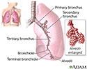
The major features of the lungs include the bronchi, the bronchioles and the alveoli. The alveoli are the microscopic blood vessel-lined sacks in which oxygen and carbon dioxide gas are exchanged.
Lungs
illustration
Aortic rupture (a tear in the aorta, which is the major artery coming from the heart) can be seen on a chest X-ray. In this case, it was caused by a traumatic perforation of the thoracic aorta. This is how the X-ray appears when the chest is full of blood (right-sided hemothorax) seen here as cloudiness on the left side of the picture.
Aortic rupture - chest X-ray
illustration
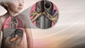
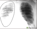
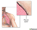
Pneumothorax occurs when air leaks from inside of the lung to the space between the lung and the chest wall. The lung then collapses. The dark side of the chest (right side of the picture) is filled with air that is outside of the lung tissue.
Pneumothorax - chest X-ray
illustration
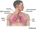
Air is breathed in through the nasal passageways, travels through the trachea and bronchi to the lungs.
Respiratory system
illustration
Chest tube insertion - series - Pleural cavity
Presentation
Pneumothorax - series - normal anatomy
Presentation

 and the chest cavity. </p>")
, bleeding into the chest (hemothorax), after surgery or trauma in the chest (pneumothorax or hemothorax), lung abscesses or pus in the chest (empyema). </p>")
. The patient may also be sedated. The chest tube is inserted between the ribs into the chest and is connected to a bottle or canister that contains sterile water. Suction is attached to the system to encourage drainage. A stitch (suture) and adhesive tape is used to keep the tube in place. The chest tube usually remains in place until the X-rays show that all the blood, fluid, or air has drained from the chest and the lung has fully re-expanded. When the chest tube is no longer needed, it can be easily removed, usually without the need for medications to sedate or numb the patient. Medications may be used to prevent or treat infection (antibiotics).</p>")


The major features of the lungs include the bronchi, the bronchioles and the alveoli. The alveoli are the microscopic blood vessel-lined sacks in which oxygen and carbon dioxide gas are exchanged.
Lungs
illustration
Aortic rupture (a tear in the aorta, which is the major artery coming from the heart) can be seen on a chest X-ray. In this case, it was caused by a traumatic perforation of the thoracic aorta. This is how the X-ray appears when the chest is full of blood (right-sided hemothorax) seen here as cloudiness on the left side of the picture.
Aortic rupture - chest X-ray
illustration
Pneumothorax occurs when air leaks from inside of the lung to the space between the lung and the chest wall. The lung then collapses. The dark side of the chest (right side of the picture) is filled with air that is outside of the lung tissue.
Pneumothorax - chest X-ray
illustration
Air is breathed in through the nasal passageways, travels through the trachea and bronchi to the lungs.
Respiratory system
illustration
Chest tube insertion - series - Pleural cavity
Presentation
Pneumothorax - series - normal anatomy
Presentation
Review Date: 1/2/2023
Reviewed By: Jesse Borke, MD, CPE, FAAEM, FACEP, Attending Physician at Kaiser Permanente, Orange County, CA. Also reviewed by David C. Dugdale, MD, Medical Director, Brenda Conaway, Editorial Director, and the A.D.A.M. Editorial team.
 All rights reserved.