
Kidney stones - InDepth
Calcium stones - InDepth; Urolithiasis Renal calculi - InDepth; Nephrolithiasis - InDepth; Stones - kidney - InDepth; Calcium oxalate - stones - InDepth; Cystine - stones - InDepth; Struvite - stones - InDepth; Uric acid - stones - InDepth; Urinary lithiasis - InDepthAn in-depth report on the causes, diagnosis, treatment, and prevention of kidney stones.
Highlights
Overview
- Kidney stones are hard, solid particles that form in the urinary tract. Stones may be as small as a grain of sand, or as large as a pea or pearl.
- If a stone (even a small one) blocks the flow of urine, excruciating pain may result, and prompt medical treatment may be needed.
- Across the globe, the incidence of kidney stones is rising, particularly in women of increasing age. It affects about 9%, or 1 in 11 people in the United States.
Risks
- Obesity and diabetes increase the risk for kidney stones.
- Dietary factors and water intake can affect your risk of developing a kidney stone.
- Certain medications such as protease inhibitors, antibiotics, diuretics, and others can increase the risk for kidney stone formation. Changes in medication may be part of an overall treatment plan.
- Taking calcium supplements beyond recommended levels may increase the risk for kidney stones.
- It is well established that drinking plenty of water helps to prevent stone formation in those susceptible and to treat kidney stones already present. The effects of other beverages, such as juice, tea, coffee, soda, and sports drinks on kidney stone formation depend on the type of stones. More research is needed.
Treatment
- Painful kidney stones require treatment. Depending on the type of stone, specific dietary changes, weight management, exercise, increased water intake, medicines, and other noninvasive treatments are available to help small stones pass through the urine.
- Large stones, stones that are causing damage to the kidneys, or stones that do not pass on their own, usually respond well to treatments such as ureteroscopy, percutaneous nephrolithotomy (PCNL), extracorporeal shockwave lithotripsy (ESWL), and laparoscopic or robotic-assisted stone surgery.
Prevention
An assessment of diet and over-the-counter vitamins and supplements usage can be useful in determining dietary recommendations that may be helpful in preventing kidney stones from recurring.
Preventive drug therapy is matched to the exact type of stone formation. For some types of stones, this can be highly effective.
Introduction
Kidney stones are hard, solid particles that form in the urinary tract. In many cases, the stones are very small and can pass out of the body without any problems. However, if a stone (even a small one) blocks the flow of urine, excruciating pain may result, and prompt medical treatment may be needed.
Urine is formed in the kidneys. The two kidneys are located deep behind the abdominal organs, below the ribs and slightly toward the outer sides of the back. About one-fourth of all the blood pumped out by the heart every minute passes through the kidneys. The kidneys filter out fluids and waste from this blood, producing urine. Kidneys remove the excess fluid, while at the same time saving necessary substances like proteins and electrolytes, and maintaining a healthy balance of salts and a normal pH (acidity level). As the urine passes through the kidneys, it becomes more concentrated. From the kidneys, urine flows through thin tubes called ureters into the bladder. The bladder's stretchy walls expand to store the incoming urine until it leaves the body through a tube called the urethra.
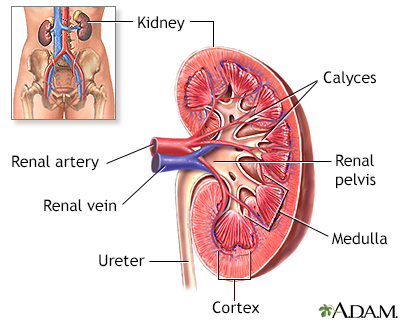
Kidney anatomy
The kidneys are responsible for removing wastes from the body, regulating electrolyte balance and blood pressure, and the stimulation of red blood cell production.
The kidneys are responsible for removing wastes from the body, regulating electrolyte balance and blood pressure, and stimulating red blood cell production.

Male urinary tract
The male and female urinary tracts are relatively the same except for the length of the urethra.
Types of Kidney Stones (Renal Calculi)
Occasionally, high levels of certain chemicals in the urine form into crystals. Eventually these crystals become large enough to form stones in the kidney, a condition called nephrolithiasis. Stones (calculi) may also form or be found in the ureter or the bladder. Combinations of minerals and other chemicals make up the salts in these stones.

Nephrolithiasis
Kidney stones result when urine becomes too concentrated and substances in the urine crystalize to form stones. Symptoms arise when the stones begin to move down the ureter causing intense pain. Kidney stones may form in the pelvis or calyces of the kidney or in the ureter.
Calcium Stones
About 80% of all kidney stones contain calcium, usually combined with oxalate, or oxalic acid. Many common foods contain oxalate.
A majority of kidney stones are composed of calcium oxalate. A smaller percentage of calcium stones are made of calcium phosphate (called brushite).
Uric Acid Stones
Uric acid is responsible for almost 10% of kidney stones. It is the breakdown product of purines, nitrogen-containing compounds found in the body and in certain foods. Uric acid enters the bloodstream, and then passes primarily into the kidneys. From the kidneys, uric acid leaves the body in the urine. Often, uric acid stones occur together with calcium stones.
Struvite Stones
Struvite stones are made of magnesium ammonium phosphate. They are almost always associated with certain urinary tract infections. Worldwide, they account for up to 30% of all kidney stones. In the United States, however, up to 15% of all stones are struvite. Most struvite stones occur in women. The rate of these stones may be declining in the United States, perhaps because of better control of urinary tract infections.
Cystine Stones
A buildup of the amino acid cystine, a building block of protein, causes 1% of kidney stones in adults and up to 8% of stones in children. The tendency to form these stones is inherited. Cystine stones grow rapidly and tend to recur. If not treated promptly, they can eventually lead to kidney failure.
Xanthine Stones
Other kidney stones are composed of xanthine, a nitrogen-containing compound. These stones are extremely uncommon and usually occur as a result of a rare genetic disorder.
Causes
The key process in the development of kidney stones is supersaturation.
- The urine carries chemicals, including calcium oxalate, uric acid, cystine, or xanthine.
- These substances can become extremely concentrated if there is not enough urine, or if unusually high amounts of crystal-forming salts are present.
- When the chemical concentration levels reach the point at which they no longer dissolve in urine, these substances form crystals.
Different factors may reduce the volume of urine, or increase the amount of stone-forming chemicals.
Deficiencies in Protective Factors
Normally, urine contains substances that protect against stone formation, including:
- Magnesium
- Citrate
- Pyrophosphate
- Various proteins and other macromolecules including enzymes, osteopontin, and glycosaminoglycans
These substances carry out the following protective actions:
- Allow chemicals in the urine to be at high concentrations without forming crystals
- Prevent crystal formation
- Coat the crystals and prevent them from sticking to the surface of kidney tubes
Not having enough of these protective substances can cause stones.
Changes in Urine Acidity
The acidity of urine is indicated by the pH scale in which a pH value of 7.0 is neutral, a low pH (below 7.0) is acidic, and a high pH (above 7.0) is alkaline. The normal pH range of urine is 4.6 to 8.0. Changes in the acid balance of the urine can affect stone formation.
- Uric acid and cystine stones mainly form in acidic urine.
- Calcium phosphate and struvite stones increase in alkaline urine.
Mixed Causes of Calcium Stones
Often, the cause of calcium stones is not known and is referred to as idiopathic (of unknown cause) nephrolithiasis. Research suggests that nearly all stones result from problems in the breakdown and absorption of calcium and oxalate. Genetic factors may play a role in about one-half of these cases. Many medical conditions and drugs can also affect digestion and intestinal absorption.
Excess Calcium in the Urine (Hypercalciuria)
Hypercalciuria is a condition in which there is too much calcium in the urine. It is responsible for as many as 70% of calcium-containing stones. A number of conditions may produce hypercalciuria. Many are due to genetic factors, but most cases are due to unknown causes (idiopathic).
The following can lead to hypercalciuria and calcium stones:
- Too much calcium absorbed by the intestines. This is usually caused by genetic factors.
- High intake of calcium, often in the form of supplements taken beyond recommended levels.
- Renal calcium leak. In this condition, the kidney does not regulate minerals normally, causing an increase of calcium in the urine.
- Increase in levels of 1,25 dihydroxy vitamin D (calcitriol, an active metabolite of vitamin D) or in the activity of the vitamin D receptor.
- Excessive sodium. High urinary levels of sodium result in increased levels of calcium. Certain defects in the kidney tubules transport system cause imbalances in sodium and phosphate, which can lead to high calcium levels in the urine. A high-salt diet can also produce this effect.
- High intake of refined carbohydrates.
- Excessive caffeine or alcohol intake.
Excess Oxalate in the Urine (Hyperoxaluria)
Oxalate is the most common stone-forming compound. Too much oxalate in the urine is responsible for up to 60% of calcium stones and is a more common cause of stones than excess calcium in the urine.
Hyperoxaluria can be either a primary or secondary condition.
- Primary hyperoxaluria is an inherited disorder in which too much oxalate in the urine is the main problem. Primary hyperoxaluria is caused by an enzyme deficiency. Genetic testing is necessary to diagnose the condition.
- Secondary hyperoxaluria results from specific gastrointestinal conditions or dietary factors that cause high levels of urinary oxalate.
Secondary hyperoxaluria is usually caused by too much dietary oxalates (found in a number of common vegetables, fruits, and grains) or by problems in the body's breakdown of oxalates. Such defects may be due to various factors:
- When fats and nutrients are not absorbed properly, calcium may bind to the unabsorbed fat instead of oxalates, which cause a buildup of oxalate. This may be due to surgery, inflammatory bowel disease, or medications.
- Severe vitamin B6 deficiencies (usually due to genetic disorders)
- Deficiencies in Oxalobacter formigenes, an intestinal bacteria that breaks down oxalate.
- Low calcium intake or low calcium absorption. When calcium is taken or eaten without other foods at mealtime, absorption may be lower.
- Excessive intake of certain over-the-counter supplements, such as vitamin C, and possibly cinnamon, cranberry, and turmeric.
- Androgens (male hormones)
Female hormones (estrogens) actually lower the risk of developing hyperoxaluria. Estrogen may help prevent the formation of calcium oxalate stones by keeping urine alkaline, and by raising protective citrate levels.
People who undergo the most common type of gastric bypass surgery, the Roux-en-Y, may be at increased risk for calcium oxalate kidney stones beginning 6 months after surgery. The added kidney stone risk is thought to be due to changes in the urine. People who have undergone Roux-en-Y gastric bypass surgery have excess oxalate and low levels of citrate in their urine after the procedure.
Gastric banding, another type of weight loss surgery, does not seem to increase the risk for kidney stones.
Excessive Calcium in the Bloodstream (Hypercalcemia)
Hypercalcemia generally occurs when bones break down and release too much calcium into the bloodstream. This is a process called resorption. It can result from several different diseases and events, including:
- Hyperparathyroidism. Overactive parathyroid glands cause about 5% of calcium stones. People with this disorder have at least a 20% chance of developing kidney stones. Women are more likely to have this disorder than men.
- Immobilization. Lack of movement can lead to kidney stones.
- Renal tubular acidosis. This disorder causes an acid and alkaline imbalance. Renal tubular acidosis not only increases calcium levels in the bloodstream, but it also reduces protective citrate levels.
High Levels of Uric Acid in the Urine (Hyperuricosuria)
Excess uric acid in the urine occurs in 15% to 20% of people (mostly men) with calcium oxalate stones. Urate, the salt formed from uric acid, creates the center of a crystal (nidus), around which calcium oxalate crystals form and grow. Such stones tend to be severe and recurrent. They appear to be strongly related to a high intake of protein. Excess uric acid in the urine also plays a major role in some uric acid stones.
Low Urine Levels of Citrate (Hypocitraturia)
Citrate is the main substance in the body that is responsible for removing excess calcium. It also blocks the process that turns calcium crystals into stones. Low level of citrate in urine is a significant risk factor for calcium stones and also increases the risk for uric acid stones. This condition most likely contributes to about one-third of all kidney stones.
Many conditions can reduce citrate levels, including:
- Too much salt in the diet
- Overweight and obesity
- Renal tubular acidosis
- Potassium or magnesium deficiency
- Urinary tract infection
- Kidney failure
- Chronic diarrhea
Low Levels of Other Stone-Blocking Compounds
Several other compounds in the urine, including magnesium and pyrophosphate, also prevent the formation of calcium stones. If any of these compounds are lacking, stones may develop.
Causes of Uric Acid Stones
Human body tissues, and certain foods, contain compounds called purines. Purine-containing foods include dried beans, peas, and liver. When the body breaks down purines, it produces uric acid. The presence of a certain level of uric acid in the body is normal, but excess uric acid can lead to stones.
The following conditions are usually seen in patients with uric acid stones:
- Too much uric acid in the urine (hyperuricosuria) for a long period of time (the most important cause of uric acid stones)
- Lower-than-normal amounts of urine produced
- Hyperuricosuria, a metabolic disorder that leads to high levels of uric acid in the urine
Note: Hyperuricosuria can also trigger calcium stones. Therefore, people with hyperuricosuria may have a combination of calcium and uric acid stones.
Many conditions and other factors may contribute to, or cause, uric acid stones:
- Gout. Uric acid and other kidney stones develop in up to 25% of people with primary gout, a painful form of arthritis that occurs when uric acid in the blood forms crystals in one or more joints.
- Diabetes. People with type 2 diabetes have highly acidic urine that can lead to kidney stones, particularly uric acid stones.
- Insulin resistance. People with insulin resistance have an increased risk for uric acid stones. The reason is unknown, but it may be related to the transport of certain salts through the kidneys. This transport changes in people with insulin resistance.
- Kidney abnormalities. Kidney problems that reduce the production of ammonia, particularly in people with diabetes or insulin resistance, may lead to the formation of uric acid stones.
- Genetic factors. Inherited factors can increase a person's risk for uric acid stones.
- Hypocitraturia. Hypocitraturia is a low amount of citrate in the urine.
- Diet. Eating too much animal protein increases the risk of forming uric acid stones.
Other risk factors include:
- Certain medications (such as chemotherapy drugs, diuretics, and salicylates)
- Binge drinking
- Not eating for long periods of time (fasting)
- Lead poisoning
- Treatment for blood cancers (such as leukemia, multiple myeloma, and lymphomas)
- Some rare types of anemia (low levels of red blood cells in the blood)
- Chronic diarrhea
Causes of Struvite Stones
Struvite stones are almost always caused by urinary tract infections. Certain bacteria produce urease, which breaks down urate and raises the concentration of ammonia in the urine. Ammonia makes up the crystals that form struvite stones. The bacteria that promote stone formation are most often Proteus, but they may also include Ureaplasma urealyticum, as well as Pseudomonas, Klebsiella, Providencia, Serratia, and Staphylococcus species. Women are twice as likely to have struvite stones as men.
Causes of Other Stones
Other stones, including cystine and xanthine stones, are usually due to genetic abnormalities.
Causes of Cystine Stones
Cystine stones develop from genetic defects that cause the abnormal transport of amino acids in the kidney and gastrointestinal system, leading to a buildup of cystine. Researchers have identified two genes responsible for this condition, which are SLC3A1 and SLC7A9.
Causes of Xanthine Stones
In some cases, xanthine stones may develop in people being treated with allopurinol for gout.
Risk Factors
Kidney stones are one of the most common disorders of the urinary tract. They are an ancient health problem. Evidence of kidney stones has been found in an Egyptian mummy estimated to be more than 7,000 years old.
Kidney stones affect more than 9% of Americans, and the prevalence of kidney stones in the United States has increased since the 1970s.
Chronic Dehydration
Not drinking enough water can increase the risk of developing a kidney stone. The amount of fluids you need can depend on the climate you live in, your activity level, and any history of kidney stones.
Gender and Age
Men
Kidney stones are two times more common in men than women, but the likelihood of stones is increasing in both men and women. The risk of developing kidney stones increases in men in their 40s and continues to rise until age 70. By age 70, 11% of men will have had a kidney stone that causes symptoms. White men have a higher risk than other ethnic groups.
Women
The risk of developing kidney stones peaks in women in their 50s. In younger women, stones are more likely to develop during the late stages of pregnancy. Pregnant women tend to have a higher calcium intake. But their kidneys do not handle the calcium as well as they did before pregnancy. Pregnant women tend to form calcium phosphate stones. Kidney stones are still rare during pregnancy, however, affecting only 1 in 1,500 pregnancies.
Risk Factors in Children
Stones in the urinary tract in children are usually due to genetic factors. Most of the time, the cause is too much calcium in the urine. Deformities in the urinary tract pose a significant risk for kidney stones in children. Babies born with a low birth weight who need to be fed intravenously are also at risk for stones.
Rates of kidney stones in adolescents have doubled in the last 25 years. Although researchers do not know the exact reason for this increase, they think it may have to do with bodyweight, diabetes and hypertension, which have been rising in young people over the years.
Obesity and Weight Gain
Obesity and weight gain are both associated with an increased risk for kidney stones.
Higher BMIs and larger waist circumferences are both risk factors for kidney stones. People with larger body sizes may excrete more calcium and uric acid into the urine, which increases the risk for kidney stone formation.
Family History
Having a family history of kidney stones doubles a person's risk for the condition. Researchers are looking into genetic markers or other factors that might predict the risk of developing kidney stones in relatives, although none has yet been clearly identified. A family history of gout may also make a person more vulnerable to developing stones.
Ethnicity
In the United States, white people seem to have the highest incidence of kidney stones, followed by Hispanics. African Americans have the lowest risk. White people are 3 times more likely to form kidney stones than African Americans. However, dietary factors can diminish any protective effects of ethnicity.
Geographical Differences
Dietary factors, minerals in local water, or climate may contribute to geographical differences that have been observed in the occurrence of kidney stones. Studies have reported the highest occurrence of kidney stones in the southern region of the United States and the lowest occurrence in the western region.
Lifestyle Factors
Specific Foods
In general, certain foods increase the risk for stones, but only in people who have a genetic or medical vulnerability. People whose diets are high in animal protein and low in fiber and fluids may be at higher risk for stones. A number of foods contain oxalic acid, but there is no proof that such foods make any major contribution to calcium oxalate stones in people who do not have other risk factors. However, several studies have shown that increasing dietary calcium and restricting salt, animal protein, and foods rich in oxalate can help prevent calcium oxalate stones from returning.
Eating a high-calcium diet does not appear to increase the risk for kidney stones, as long as the diet also contains plenty of fluids, potassium, and phosphate. (Increasing calcium alone may pose a modest risk for stones.)
Calcium supplements on the other hand have been associated with an increased risk for kidney stones in several studies. For example, the Women's Health Initiative showed that postmenopausal women who took 1,000 mg calcium/400 I.U. vitamin D supplements daily had a 17% higher risk of developing kidney stones. Calcium from food sources appears to be the best option.
Stress
People who have had a major, stressful life experience may be more likely to develop stones. Some experts speculate that this increased risk may be due to a hormone called vasopressin, which is released in response to stress. Vasopressin also decreases the volume of urine, which makes the chemicals in urine more concentrated and prone to forming crystals and stones.
Being Bedridden
Any medical or physical condition that keeps a person in bed or immobile increases blood levels of calcium from bone breakdown, thereby posing a risk for stone formation.
Medical Conditions
Diabetes
People with type 2 diabetes are at increased risk of developing kidney stones, particularly uric acid stones.
Gout
People with gout are at high risk for uric acid stones.
High Blood Pressure
People with high blood pressure are up to three times more likely to develop kidney stones. It is not entirely clear whether having high blood pressure increases the risk for a stone, or if stones lead to high blood pressure, or if there is a factor linking both conditions.
Inflammatory Bowel Disease (IBD)
Crohn disease and ulcerative colitis cause problems in the absorption of substances in the intestines. These problems significantly increase the risk for kidney stones, particularly in men.
Urinary Tract Infections (UTIs)
UTIs are almost always the cause of struvite stones.
Hyperparathyroidism
The parathyroid glands regulate calcium levels in the body through parathyroid hormone. In hyperparathyroidism, one or more of these glands makes too much parathyroid hormone. Some people with hyperparathyroidism develop kidney stones. Surgery to remove the hyperactive parathyroid gland in such patients reduces the risk for stone formation, but the risk still remains high for some time after surgery.
Other Medical Conditions
Kidney disease, chronic diarrhea, other GI conditions such as celiac disease or surgery in the intestines, certain cancers (such as leukemia and lymphoma), and sarcoidosis (a type of systemic disorder that causes inflammation and release of parathyroid hormone-like protein) put people at higher risk for stones.
Medications
Certain Gout Medications
Some medicines used to treat gout act to lower uric acid levels in the blood by encouraging the kidneys to get rid of more uric acid. As a result, higher levels of uric acid end up in the urine (hyperuricosuria), and can lead to an increased risk for kidney stones. These agents should not be used for individuals who already have higher urine levels of uric acid.
AIDS medications
More than 10% of AIDS patients who take the medicine indinavir develop stones. The risk is even higher in people with AIDS who also have hepatitis B, hepatitis C, or hemophilia, as well as those who are very thin or who take the antibiotic combination of trimethoprim/sulfamethoxazole (TMP-SMX).
Other Drugs
Kidney stones are a rare side effect of thyroid hormones and loop diuretics (drugs that increase urination), although diuretics are also used to prevent calcium stones. Certain cancer chemotherapy drugs can also cause kidney stones. Long-term use of medications such as calcium-based antacids, which change the acid content of urine, may increase the risk for kidney stones.
Ephedrine (found in some drugs used to treat asthma and congestion), triamterene (sometimes used to treat high blood pressure and fluid build-up), magnesium antacids (silicates), TMP-SMX (used to treat certain infections), topiramate (used to treat certain seizures), carbonic anhydrase inhibitors (used to treat glaucoma), furosemide (used to treat fluid retention), vitamin C or D excess, and overusing laxatives may also promote the formation of kidney stones.
Certain antibiotics and drug therapies for type 2 diabetes are also associated with kidney stone formation.
Symptoms
In many cases, kidney stones do not produce symptoms. However, if a stone becomes trapped (or lodged) in the ureter (the thin tube between the bladder and kidney), symptoms can be very severe. Often, symptoms vary depending on the stone's location and its progress.
Kidney stone attacks (renal colics) are most common late at night or in the early morning, possibly because of low urine output during these times. A renal colic is typically characterized by sudden and extreme flank pain.
- Pain usually begins abruptly on one side and then continues to be intense and consistent. In some cases, it lasts for a few minutes, disappears, and then returns after about 10 minutes.
- The person cannot find a comfortable position and usually stands, sits, paces, or reclines in a failed search for a position that will bring relief.
- If the stone is in the kidney or upper urinary tract, the pain usually starts in one flank area (to the side of the upper back). It typically radiates to the groin.
- If the stone is too large to pass easily, the pain follows the muscle contractions in the wall of the ureter as they try to squeeze the stone along into the bladder.
- Nausea and vomiting may occur.
- Blood may be present in the urine.
- As the stone passes down the ureter, closer to the bladder, the person may feel the need to urinate more often or a burning sensation during urination. The location of the pain may shift to the lower abdomen on the side of the stone.
- A fever suggests that the person may also have a urinary tract infection.
The size of the stone does not necessarily predict the severity of the pain. A very tiny crystal with sharp edges can cause intense pain, while a larger round stone may not be as distressing. Struvite stones can often occur without symptoms.
Diagnosis
The doctor will perform a physical exam. This includes tapping on the back over the kidneys and pressing on the abdomen to detect tender locations.
Medical History
The person's age is a significant factor in the diagnosis. Kidney stones that occur in children and young people are more likely to result from inherited problems that cause excess cystine, xanthine, or, in some cases, calcium oxalate. In adult people, calcium stones are most common.
A medical history may help predict which crystal has formed the stone. The doctor will need to know the following:
- Any previous kidney stone attacks
- History of cancer, sarcoidosis, or small bowel disease
- Any medications being taken, including nonprescription substances, particularly high doses of vitamins D or C and calcium-containing antacids
Ruling Out Other Disorders
Many conditions can cause symptoms similar to those of kidney stones. Usually the diagnosis is easily made because of the type of symptoms, but it is not always clear. Urinary tract infections can cause similar, but usually less intense, pain. In fact, people with an infection may also have a kidney stone.
Other causes of pain that may mimic kidney stones include:
- Gallstones
- Diverticulitis (infection or irritation of abnormal pouches in the intestines)
- Intestinal blockage
- Blood clots
- Irritable bowel syndrome (IBS)
- Appendicitis
- Peptic ulcers
- Pancreatitis (inflammation of the pancreas)
- Hepatitis
- Pelvic inflammatory disease
- Inflammatory bowel disease (Crohn and colitis)
- Heart attack
- Pyelonephritis (a kidney infection)
- Blockage of urinary flow down the ureter due to a scar, tumor or congenital defect
Imaging Techniques
Various imaging techniques are helpful in determining the presence of kidney stones. The best approach uses spiral (or helical) computed tomography (CT) scans. If these scans are not available, the person will need an ultrasound or standard x-rays. If no stones show up, but the person has severe pain that suggests the presence of kidney stones, the next step is an intravenous pyelogram (IVP).
X-Rays
A standard x-ray of the kidneys, ureters, and bladder may be a good first step for identifying stones, because about 80% of stones are visible on x-rays. Calcium stones can be identified on x-rays by their white color. Cystine crystals can also show up on x-rays. X-rays may not provide enough detail about the exact location of a stone.
Computed Tomography (CT)
CT scans use x-rays with computer technology to produce 2D images of the body. A type of CT scan called a spiral or helical CT scan is currently the most accurate method for diagnosing stones in either the kidneys or the ureters. CT scans can be used to find the exact location of the stone, as well as to look for anatomical problems that may have led to stone formation. This test provides detailed, accurate images of even very small stones. A spiral CT scan can often identify other causes of pain in the kidney area. However, radiation exposure is an issue with CT scans and overuse is a concern.
Ultrasound
Ultrasound is a noninvasive technique that uses sound waves to create images of internal organs and structures. Ultrasound is now often used as the initial screening test for stones. It is also used to screen patients who need monitoring for stone recurrence. It is not useful for finding very small stones, but some research indicates that it may be an effective first diagnostic step in the emergency room to help identify whether a person has a stone. Ultrasound will not help identify stones that are in the ureter but can help determine if there is a blockage to the flow of urine out of the kidney. Ultrasound is also effective in children.
Intravenous Pyelogram (IVP)
With IVP, the doctor injects a special dye into the person's vein. A technician then takes x-rays as the dye enters the kidneys and travels down the urinary tract. The dye helps visualize any abnormalities in the urinary system. Where it is available, spiral CT is now preferred, since it gives a faster diagnosis and is more accurate, safer, and similar in cost.
IVP should not be used on patients with kidney failure. There is also a risk for an allergic reaction to standard dyes, although newer, less allergenic dyes are becoming available.

Intravenous pyelogram (IVP)
In the procedure intravenous pyelogram (IVP), the patient is injected with radiopaque dye and X-rays are taken as the dye travels through the urinary tract. This procedure is performed to confirm the presence of kidney stones, although some stones may be too small to see.
In the procedure intravenous pyelogram (IVP), the person is injected with dye. X-rays are taken as the dye travels through the urinary tract. This procedure is done to confirm the presence of kidney stones, although some stones may be too small to see.
Magnetic Resonance Imaging (MRI)
MRI uses powerful magnets to provide detailed images of internal organs and tissues. MRI techniques are showing promise for diagnosing urinary tract obstruction, but they do not yet accurately reveal small stones, or stones that do not cause a blockage. Because no radiation is involved with MRIs and ultrasounds, they are good options for children and pregnant women.
Urine Tests
Urine samples are needed to evaluate features of the urine, including its acidity and the presence of:
- Red or white blood cells
- Infection
- Crystals
- High or low levels of chemicals that inhibit or promote stone formation
Clean-Catch Urine Sample for Culturing
After determining that a kidney stone is present, the health care provider usually gives the patient a collection kit, including filters, to try to catch the stone or gravel as it passes out. The type of crystals that make up the stone can be determined (crystallography). The urine may also be tested (cultured) for the presence of infection-causing organisms. A clean-catch urine sample is almost always required for culturing. To provide a clean catch, do the following:
- First, wash your hands thoroughly, and then wash the penis or vulva and surrounding area 4 times with downward strokes, using a new soapy sponge each time.
- Begin urinating into the toilet and stop after an ounce or two.
- Position the container to catch the middle portion of the urine stream.
- Urinate the remainder into the toilet.
- Tighten the cap on the container securely, being careful not to touch the inside of the rim.

Calcium urine test
During a calcium urine test, urine is collected for a 24-hour period. The urine will later be analyzed for the amount of calcium content.
Twenty-Four Hour Urine Collection
A 24-hour urine collection may be needed to measure urine volume and levels of acidity, calcium, sodium, uric acid, oxalate, citrate, and creatinine.
- You should not change any of your usual eating or drinking patterns when performing this test.
- Discard the first urination on the day of the test.
- Afterward, collect all urine passed over the next 24 hours, including the first urination on the morning of the second day.
- A second 24-hour urine collection may be needed to determine whether treatment is working, or it may be done if the first analysis was not conclusive and the doctor suspects a less common stone, such as a cystine or xanthine stone.

Uric acid test
Uric acid urine test is performed to check for the amount of uric acid in urine. Urine is collected over a 24 hour period and is sent to the laboratory for testing. The most common reason for measuring uric acid levels is in the diagnosis or treatment of gout, because gout results from crystallization of uric acid crystals in joints. Some kidney stones are also composed of uric acid.
Testing Urine Acidity
Testing whether urine is acidic or alkaline helps to identify the specific type of stone. The levels of acidity or alkalinity in any solution, including urine, are indicated by the pH scale:
- A pH value of 7.0 is neutral.
- A solution with a low pH (below 7.0) is acidic. (A low pH favors the development of uric acid and cystine stones.)
- A solution with a high pH is alkaline. (A high pH favors the development of calcium phosphate and struvite stones.)
Urine tests that are used to determine the specific chemical and biological factors causing the stone should be performed about 6 weeks after the attack, since the attack itself may change the levels of such substances, including calcium, phosphate, and citrate.
Note that calcium levels in the urine may be abnormal even in many people who do not have stones. In addition, high urinary concentrations of calcium may pose a greater or lesser risk for stones, depending on a person's age.
Testing for Blood in the Urine
A dipstick test for blood in the urine (called hematuria) is typically performed when people come to the emergency room with flank pain (the primary symptom of kidney stones). About one-third of people with kidney stones do not show blood in the urine, so other tests may be needed.
Blood Tests
Blood Tests for Stone Factors
Blood and urine tests help determine what substances formed the crystals. This allows the doctor to determine the appropriate treatment and preventive measures.
Blood tests may help determine blood levels of urea nitrogen, creatinine, calcium, phosphate, and uric acid in people with known or suspected calcium oxalate stones. Doctors will usually schedule these tests about 6 weeks after the attack, in order to measure these substances when the stone has been passed and the patient has been stabilized. This is particularly true in people with recurrent stones.
Parathyroid Tests
Tests to detect parathyroid hormone levels are given if the doctor suspects hyperparathyroidism based on other signs and symptoms.
Tests for Infection
A test result that shows a high white blood cell count might indicate infection. Such results, however, could be misleading, since the number of white blood cells could also increase in response to the extreme physical stress of a kidney stone attack.
Tests for Metabolic Problems
About one-half of children with stones have an identifiable metabolic disorder, which increases five times their risk for stone recurrence. Experts argue whether tests for metabolic disorders are routinely needed once the stone composition has been determined. Studies suggest the following:
- People with recurrent calcium stones have a wide range of irregular blood or urine test results, indicating a variety of possible metabolic disorders. For example, calcium stones in middle-aged women may be due to parathyroid abnormalities.
- Calcium phosphate stones most likely result from renal tubular acidosis.
- People with non-calcium stones generally have identifiable metabolic disorders.
- Determining the stone composition may be sufficient for treatment, and may help avoid unnecessary metabolic tests.
Microscopic Examination
Kidney stones that are removed are sent for stone analysis (crystallography).
Treatment
When tests show someone has a kidney stone, the next step is to determine treatment. People who have severe pain, vomiting, fever, or symptoms of infection should be evaluated and treated in the emergency room. The size, location, type, and number of stones are important factors in determining the best treatment.
- Any infection should be treated.
- If symptoms are severe or if the size of the stone is large enough that is not likely to pass by itself, a procedure will be done to remove the stone. This will relieve the symptoms, reduce the risk of developing infection, and avoid kidney damage.
People diagnosed with a type of stone called a staghorn stone almost always undergo procedures to remove it, in order to avoid kidney damage. These stones develop in the part of the kidney where urine collects before leaving the kidney. They are most often associated with struvite stones and infection.
For patients without the above symptoms or findings, a decision must be made regarding the need for a procedure to treat the stones. One or more of the below options may be best:
- Simple observation
- Medicines to help a stone pass
- Medications to reduce the chance of future stones
- Dietary changes to reduce the chance of future stones
Small stones that are causing no or tolerable symptoms in otherwise healthy people can be observed. Around 80% of stones less than 5 millimeters in size will pass on their own. Less than half of stones that are 5 to 10 millimeters will pass on their own.
Stones that are larger or are located closer to the or within the kidney are more likely to cause recurrent symptoms.
Treatment for Severe Attacks
Strong opioid painkillers are often required for a severe kidney stone attack. However, doctors will usually not give such drugs until they confirm the presence of a kidney stone with an imaging study.
Watchful Waiting
In about 80% of people, the kidney stones are small enough (about 5 mm or less) that they pass through normal urination, usually within 2 to 3 days. In some cases, a stone may take weeks to months to pass, although the pain usually goes away before that.
The person should drink plenty of water (2 to 3 quarts, or 1.9 to 2.85 L, a day) to help move the stone along, and take painkillers as needed. The doctor usually provides a collection kit with a filter and asks the patient to save any passed stones for testing.
If the stone has not passed in 2 to 3 days, the patient may need additional treatments depending on the level of pain or change in kidney function. In some severe cases, hospitalization may be necessary.
Alpha-blockers (such as tamsulosin) can relax muscles in the urinary tract, helping kidney stones pass.
Medications
Medications may be used to help a stone pass or to prevent the formation of new stones. The evidence supporting some of these are not strong, however.
Medications That Help Stones Pass
A type of medicine, which has other uses, may help smaller stones pass. These drugs are called Alpha-blockers and they help relax smooth muscles in the ureter and bladder, thus potentially helping a stone move.
They include terazosin (Hytrin, generic), doxazosin (Cardura, generic), tamsulosin (Flomax, generic), alfuzosin (Uroxatral), and silodosin (Rapaflo).
Medications for Calcium Stones
Diuretics
Diuretics are medicines commonly used to treat high blood pressure and other disorders. They remove fluid and sodium from the body. Low doses of a class of diuretics, known as thiazides, are sometimes used to reduce the amount of calcium the kidneys release into the urine, thereby reducing recurrent calcium stone formation. Thiazides include:
- Hydrochlorothiazide (Avalide, Hydra-zide)
- Chlorothiazide (Diuril)
- Chlorthalidone
However, thiazides also cause potassium loss, which reduces citrate levels and can increase the risk for stones. People taking thiazide pills may also take potassium citrate to prevent citrate loss. Amiloride (Midamor) is a diuretic which does not lower potassium levels and which may be used if a thiazide does not work. Thiazides may cause muscle cramps. Researchers are looking at lower dosing options for thiazides.
Citrates
Citrate salts are often given to people with calcium oxalate or uric acid stones:
- Potassium magnesium citrate is available over the counter. It is proving to be very beneficial in preventing kidney stones.
- Potassium citrate (such as Urocit-K) is the only treatment given to people with normal urine calcium levels. Between 70% and 75% of people with recurrent stones have ongoing remission (no stone recurrence) with potassium citrate treatment. However, some people cannot tolerate potassium citrate because of side effects (stomach problems).
- Magnesium citrate (such as Citroma) may help people who develop calcium stones from impaired intestinal absorption due to short bowel disease.
Citrates may cause stomach upset. People with struvite stones, urinary tract infections, bleeding disorders, or kidney damage should not use these products. People who take citrate supplements containing potassium should not take any other medications that contain potassium or prevent its loss. People with peptic ulcers should avoid citrate supplements, or discuss using non-tablet forms with their doctors.
Phosphates
Phosphates help reduce the breakdown of bone that releases calcium into the bloodstream. They are also involved in the kidney's reabsorption of calcium from the urine.
Neutral (nonacidic) sodium or potassium phosphate (such as K-Phos, Neutral, and Neutra-Phos) is usually taken 4 times a day after meals to prevent kidney stones. Diarrhea is a possible side effect.
Avoid acidic forms of phosphate, because they increase the risks for both hypocitraturia and hypercalciuria. They may cause gastrointestinal distress.
Cholestyramine(such as Questran and Questran Light)
A drug used to reduce cholesterol levels. However, because it binds with oxalate in the intestine, it is also used to reduce high oxalate levels in urine (hyperoxaluria). The drug usually comes in a powder that is dissolved in liquid. Bloating and constipation are common side effects of this drug. Cholestyramine also interferes with other medications, including digoxin (Lanoxin) and warfarin, and it may contribute to calcium loss and osteoporosis. In order to prevent such interactions, take other drugs 1 hour before, or 4 to 6 hours after taking cholestyramine.
Long-term use of cholestyramine may cause deficiencies of vitamins A, D, E, and K. Vitamin supplements may be necessary.
Medications for Uric Acid Stones
Sodium Bicarbonate
People who have uric acid stones from acidic urine may take sodium bicarbonate to reduce urine acidity. People taking sodium bicarbonate must test their urine regularly with pH paper, which turns different colors depending on whether the urine is acidic or alkaline. Too much sodium bicarbonate can cause the urine to become too alkaline. This increases the risk for calcium phosphate stones. People who need to reduce the amount of sodium they take in (as a result of other medical conditions) should not use sodium bicarbonate.
Potassium Citrate
Potassium citrate, which restores citrate to the urine, is useful for people with high levels of uric acid in the urine.
Allopurinol
Allopurinol (Lupurin, Zyloprim) is very effective at reducing high blood levels of uric acid, and it may be helpful for people with uric acid stones. Allopurinol will not prevent calcium stones from forming. There is also a slight risk for the formation of xanthine stones with this drug. Side effects include diarrhea, headache, muscle pain, and fever. About 2% of people have an allergic reaction to allopurinol that causes a rash. In rare cases, the rash can become severe and widespread enough to be life-threatening.
Allopurinol reduces uric acid levels rapidly, so it may trigger an attack of gout in vulnerable people. To relieve this problem, people taking allopurinol should also take a nonsteroidal anti-inflammatory drug (NSAID) for 2 or 3 months. Aspirin should not be taken, because it increases uric acid levels. People should discuss the appropriate NSAID choice with their doctor.
Febuxostat (Uloric) lowers uric acid levels by inhibiting the same enzyme as allopurinol. People who are allergic to allopurinol can take febuxostat. This newer drug is very effective, but expensive.
Medications for Struvite Stones
Before people can receive any medical treatment for struvite stones, they must have the stones removed with one of the techniques below.
Antibiotics for Eliminating Infection
People with struvite stones should receive ongoing treatment with antibiotics to keep the urine free of the bacteria that cause urinary tract infections. Careful follow-up and urine testing are extremely important. A high urine pH indicates low acidity and an increased risk for infection.
Acetohydroxamic Acid (AHA)
AHA (Lithostat) is beneficial when used with long-term antibiotics. AHA blocks urease, an enzyme released by bacteria which increases ammonia in urine. AHA has been effective in preventing stones even when bacteria are present. Side effects, however, can be severe. The drug reduces iron levels in the body, so anemia is a common problem. People may need to take iron supplements. Other side effects include nausea, vomiting, depression, anxiety, rash, persistent headache, and, rarely, small blood clots in the legs. This drug is recommended only for people with healthy kidneys whose condition is due to specific struvite-causing organisms. People taking this medicine should avoid alcohol. Pregnant women should not take AHA.
Organic Acids
Medical treatments to dissolve stones may be useful in people who do not respond to other medications, or in combination with surgery. Acidic urine dissolves struvite stones, so the doctor may wash the urinary tract with a solution of organic acids (such as Renacidin). Candidates for such washes must have sterile urine (no bacteria or other organisms in the urine) and healthy kidney function. In surgical patients, the wash is performed 4 or 5 days after the operation. The wash starts with saline (salt solution) for 1 to 2 days. If there are no problems, the organic acid solution follows for another 1 or 2 days, until all stones dissolve. Regular urine tests are necessary to ensure that the bacteria do not return.
Medications for Cystine Stones
The first-line treatment for cystine stones is increasing the alkalization of urine so the stones can dissolve. If alkalization fails, drug treatments may include d-penicillamine, alpha-mercaptopropionylglycine (tiopronin), or captopril. These medications lower cystine concentration.
People with cystine stones must drink much more fluid than people with other stones, at least 4 quarts (3.8 L) of water a day.
Other Treatments
Surgery is usually needed if:
- The stone is too large to pass on its own
- There are signs that the stone is growing
- The stone is blocking the urine flow
- It is causing a urinary tract infection or kidney damage
- It is a struvite stone
Almost all renal stones 1 cm or smaller may be treated with shock wave lithotripsy, ureteroscopy, or percutaneous (through the skin) nephrolithotomy.
Today, treatments for stones are much less invasive than in the past. Major surgery is performed in less than 2% of people. Stone removal procedures include the following:
- Extracorporeal shock wave lithotripsy (ESWL) is used for small stones (smaller than 2 cm, or about three-quarters of an inch) that occur in the upper part of the ureter and do not pass on their own. Slightly more than one-half of stones are less than 1 cm in diameter and are quite responsive to ESWL. Lithotripsy might even be safe and effective for people whose stones are associated with malformed kidneys, although such people are at higher risk for stone recurrence and should be carefully monitored. ESWL takes less time to perform and requires a shorter hospital stay than percutaneous nephrolithotomy.
- Ureteroscopy (URS) is generally the best procedure for stones in the lower urinary tract, although lithotripsy is also usually feasible and patients ordinarily prefer it. Ureteroscopy may still be used for some stones located in the kidney.
- Percutaneous nephrolithotomy (PCNL) can be used for large stones in the upper urinary tract, when ESWL or ureteroscopy fails, for kidney transplant patients, or when the kidneys or surrounding areas are malformed. PCNL may produce better clearance of moderate size stones (1 to 2 cm) than ESWL. PCNL is the preferred procedure for drug-resistant cystine stones, which are usually also resistant to shock wave therapy.
- Laparoscopic or robotic-assisted stone surgery may be required if these procedures fail or are not appropriate, or in special cases, such as when the personis very obese. Rarely an open procedure is needed.
Most procedures are more effective for calcium and uric acid stones and less effective for struvite and cystine stones, although new techniques may be improving their effectiveness on all stones.
Brushite (calcium phosphate) stones, calcium oxalate monohydrate, and cysteine stones are all more resistant to ESWL therapy and can be expected to have better rates at all sizes and locations with URS.
Extracorporeal Shock Wave Lithotripsy (ESWL)
Extracorporeal shock wave lithotripsy (ESWL) is a technique that uses sound waves (ultrasound) to break up simple stones in the kidney or upper urinary tract. (Extracorporeal means "outside the body," and lithotripsy means "stone-breaking.") ESWL is not used for cystine stones. The procedure generally does not work for stones larger than 2 centimeters in diameter. ESWL can often be done on an outpatient basis with limited anesthesia such as IV sedation and topical agents.
There are several variations of ESWL. The following is a typical procedure:
- The person is positioned in a water bath. (In some procedures, the person lies on a soft cushion.)
- The procedure uses ultrasound to generate shock waves that travel through the skin and body tissues until they hit the dense stones. (The doctor pinpoints the stone during treatment by using x-rays or ultrasound.)
- The shock waves crush the stones into tiny sand-like pieces that usually pass easily through the urinary tract.
The shattered stone fragments may cause discomfort as they pass through the urinary tract. If so, the doctor may insert a small tube called a stent through the bladder into the ureter to help the fragments pass. This practice, however, does not usually speed up passage of the stones and is not used routinely.

Lithotripsy procedure
Extracorporeal shock wave lithotripsy (ESWL) is a procedure used to shatter simple stones in the kidney or upper urinary tract. Ultrasonic waves are passed through the body until they strike the dense stones. Pulses of sonic waves pulverize the stones, which are then more easily passed through the ureter and out of the body in the urine.
Extracorporeal SWL is a procedure used to shatter simple stones in the kidney or upper urinary tract. Ultrasonic waves are passed through the body until they strike the dense stones. Pulses of sonic waves pulverize the stones, which then pass more easily through the ureter and out of the body in the urine.
This method cannot be performed for patients who are pregnant, have bleeding risks, or have an untreated urinary tract infection.
ESWL has a 50% to 90% success rate, depending on the location of the stone, the size of the stone, and the surgeon's technique and experience. It is most effective for stones 1 cm or smaller. Stones sized 1 to 2 cm may need extra treatment sessions. ESWL is not as effective as percutaneous nephrolithotomy (PCNL) for stones in the lower urinary tract, but it sometimes reduces the length of treatment and the hospital stay. Recovery time is short. Most people can resume their normal activities in a few days.
Obesity and urinary system abnormalities may also lower how well it works.
ESWL is considered generally safe, and has few complications. If they do occur, complications may include:
- Blood in the urine, the most common complication, may last for a few days after treatment. To reduce the chances of bleeding, doctors usually tell people to avoid taking aspirin and other NSAIDs (which can promote bleeding) for 7 to 10 days before the treatment.
- Bruising and minor discomfort due to the shock waves are common in the back or abdomen.
- Sometimes the stone does not completely break up with one treatment, and additional treatments may be required. Inability to pass stone fragments may also be a particular problem in people who have cysts or other kidney problems.
- ESWL may sometimes produce kidney damage.
- Hematoma – there could be bleeding that surrounds the kidney. This usually will resolve spontaneously. Sometimes a blood transfusion may be needed.
ESWL appears to be safe for children. Experts recommend using the least amount of shocks and impact possible in young people. If more than one treatment is needed, the person should wait at least 15 days before having the next treatment.
Ureteroscopy
Ureteroscopy may be used for stones in the middle and lower ureter. Because it uses smaller instruments, this procedure can be done successfully in children.
During ureteroscopic stone removal:
- The person receives a general anesthetic, though no incision is required for the procedure.
- The surgeon passes a small fiberoptic instrument called a ureteroscope through the urethra and bladder into the ureter.
- The surgeon locates the stone or stones.
- The urologist can use a laser to break up the stones, or remove them with a basket, grabber, or suction tool.
- The surgeon may decide to leave a small tube, or stent, in the ureter for a few days after treatment, to help the lining of the ureter heal.
Ureteroscopy achieves a higher stone removal rate than ESWL, but it also has a higher risk of developing complications. In some cases, large stones are not broken up into small enough pieces. This can result in a blockage of the urinary tract and possible kidney damage.
Imaging tests, such as ultrasound or spiral CT, are useful within 3 months to check for residual stones, and a second procedure may be required. The risk of developing complications is highest when the procedure is performed by less experienced surgeons, or if stones are found in the kidney. The risk for perforation of the ureter increases the longer the procedure takes.
Percutaneous Nephrolithotomy (PCNL)
Percutaneous nephrolithotomy (PCNL) may also be used to treat kidney stones. It is often considered when ESWL is not available or when the stone is very large, in a difficult to reach location, or is a cystine stone. PCNL is preferred over ESWL for stones that have remained in the ureter for more than 4 weeks. PCNL is recommended for stones greater than 2 cm diameter and perhaps for moderate size stones as well (1 to 2 cm). PCNL is also the treatment choice for partial and complete staghorn kidney stones.
PCNL is more effective than ESWL for people who are severely obese, and it appears to be safe for the very old people and the very young. Success rates are very high for kidney stones and for ureteral stones. However, success may vary based on the technique used and the specific person. For example, success rates are slightly lower in children, although the procedure can be done safely in young people. Long-term effects are unknown.
A typical procedure is as follows:
- The surgeon makes a tiny incision in the skin of the back or flank and creates a channel directly into the kidney.
- The surgeon then inserts an instrument called a nephroscope through the channel.
- The stone is located and removed. If it is large, it is destroyed using ultrasound, lasers, or other devices. The surgeon then removes the fragments. An advantage of PCNL over ESWL is that the surgeon is able to remove the stone fragments directly, instead of relying on their natural passage from the kidney.
- Generally, people stay in the hospital for 1 to 5 days. Sometimes a person may be able to go home on the same day.
- Either a small device, called a nephrostomy tube, is left in the kidney for drainage. Or, a ureteral stent that keeps the ureter open may be used during the healing process. Both will be removed at some point after surgery. If neither is needed, the hospital stay may be shorter and there may be less pain.
Devices Used to Destroy Stone
For large stones, some type of energy-delivering device may be needed to break the stone into small pieces. These are referred to as intracorporeal lithotripsy devices (meaning stone breakers within the body), and may include:
- Ultrasound is currently the preferred method. It results in a stone-free rate of more than 90%. A rigid nephroscope delivers the ultrasound waves.
- Pneumatic (compressed air) lithotripsy uses a probe that comes in direct contact with a stone. Compressed air causes a piston to collide rapidly with the probe, and the result is a "jackhammer" action that breaks up the stone. However, this method can send stone fragments into other parts of the urinary tract.
- A more recent device uses a combination pneumatic probe and ultrasound. It produces stone-free rates of over 80%. It may prove to be superior to ultrasound alone, and it is effective against stones of all types.
- The holmium laser uses a flexible nephroscope to literally melt the stones. This laser destroys up to 100% of all types of stones and it has an excellent safety record. It should be used sparingly, however, and with particular caution on large uric acid stones until more is understood about its effect. (Another device, the erbium: YAG laser shows promise for lithotripsy, but is not currently in clinical use.)
Complications
Advances in technology have reduced the number and severity of complications related to PCNL in recent years. Bleeding during surgery is the most common complication. Post-operative bleeding occurs in about 1% of PCNLs. Tears and injury to nearby organs or structures may also occur. Scarring of the tissue is one possible complication, but studies indicate that this scarring does not impair kidney function. Up to one-third of people experience fever after PCNL.
Because the procedure uses large volumes of fluid, fluid overload is a potential problem, particularly in children or people with heart disease.
Infection may occur in some patients. Other serious complications may include a collapsed lung and injuries to areas outside the kidney (but within the operative area), such as the abdomen or chest. But these occurrences are extremely rare.
Open Surgery (Nephrolithotomy)
Open surgery is the least common method for removing stones. It is only performed in a very small percentage of people today. Called nephrolithotomy, it involves making incisions through the person's flank and into the kidney. The surgeon will cool the kidneys using ice. X-rays taken during the procedure help locate the stone. The surgeon will isolate the arteries supplying the kidneys to ensure they are not harmed during the surgery. The surgeon will then locate and remove the stone. The surgeon will also correct any blockage in the affected area.
This surgery is very invasive and therefore is only used for:
- People with very large or complex stones that cannot be removed safely using less invasive measures
- Very obese people
- People who have kidneys with anatomical abnormalities or obstructions
- People undergoing open surgery for another condition
Some centers report success with ESWL in people who would normally be nephrolithotomy candidates. Therefore, even these people should discuss other options with their surgeon and seek advice from experienced centers.
The procedure is not appropriate for people with:
- Bleeding or clotting disorders
- Untreated widespread infection
- Severe and chronic kidney disease (unless removing the stone will improve kidney function)
Complications
Effects on the Urinary Tract and Kidneys
Obstruction and Infection
Although kidney stones often lead to obstruction (blockage) of the urinary tract, the blockage is usually temporary and causes no lasting damage. In some cases, however, particularly if the obstruction progresses with no symptoms, infection may occur, which can be serious and requires immediate attention.
Chronic Kidney Disease
People with kidney stones face a higher risk for chronic kidney disease, particularly if they also have diabetes, high blood pressure, or frequent urinary tract infections. Having chronic kidney disease, in turn, can increase the risk for a heart attack. Researchers have found that in general, kidney stones increase the risk for a heart attack by more than one third (as well as the risk for certain other disorders), independent of their effect on chronic kidney disease.
Kidney Failure
It is very rare for kidney stones to cause kidney failure. However, some people have risk factors that make them more vulnerable to this serious complication, such as:
- Very frequent stone recurrences (such as in people with cystine stones or other inherited forms of kidney stone disorders)
- Episodes of urinary tract infections with obstruction, a particular risk with struvite stones
- A history of having many procedures for kidney stones
- Greater size of the kidney stone gravel
Long-Term Outlook: Risk for Recurrence
Without treatment, calcium stones recur in 40% of people within 5 years of the first attack, and in 75% of people within 20 years. The individual risk for recurrence, however, varies depending on the stone and the underlying condition. For example, a 15-year-old with inherited cystine stones has a very high risk for recurrence, while a middle-aged man with a first calcium oxalate stone has a good chance of never passing another stone.
Prevention
Anyone who has had kidney stones should try to prevent a recurrence. Some general prevention tips include:
- Increase fluid intake, restrict sodium, and reduce protein intake
- Get more potassium
- Try to correct any dietary habits that cause acid or alkaline imbalances in the urine, which promotes stone formation
Dietary Considerations
People with kidney stones appear to be more sensitive to certain foods than people who do not form kidney stones. Therefore, vulnerable people should make specific changes in their diet.
Because different kidney stone types may require specific dietary changes, patients should work with their doctors to develop an individualized plan. Tailored nutritional considerations are very important in preventing recurrences, and people should comply with the proper diet. Drinking plenty of fluids is important for preventing a recurrence of any kidney stone.
Indications for Drug Treatments
If dietary treatments fail, drug therapy may be helpful. A number of drugs are available to prevent recurrences of calcium oxalate and other stones. Medications that inhibit the formation of stones in some cases include allopurinol, thiazides, potassium citrate, and potassium-magnesium citrate. In addition, drug treatments can sometimes help prevent other complications related to stones, such as osteoporosis.
Correcting Underlying Conditions Known to Cause Kidney Stones
It is also important to treat and correct, if possible, any underlying disorder that may be causing stones to form. Such disorders include distal renal tubular acidosis, hyperthyroidism, sarcoidosis, and certain cancers. To prevent calcium stones that form in hyperparathyroid patients, a surgeon may remove the affected parathyroid gland (located in the neck).
Fluids (Water, Juice, and Other Beverages)
Of all the preventive recommendations, drinking enough fluids is the most important guideline for people with any type of kidney stones.
- In general, people with calcium or uric acid stones should drink at least 10 full glasses (2.4 L) of fluid each day (at least one half should be water). They should drink one glass with each meal and drink fluids at night, even if they have to get up during the night to go to the bathroom. Fluid intake should produce at least two-and-a-half quarts (2.4 L) of urine each day.
- To prevent cystine stones, people should drink even more water, more than a gallon, or sixteen 8-ounce cups (3.8 L), every day. People should drink this amount at regular intervals throughout the day and night.
In all cases, people need more fluid after exertion and during times of stress. If they drink enough, the urine should be pale and almost watery, not dark and yellow.
Water
Water hardness (meaning how much calcium is in the water) generally plays only a small role in stone formation. By far, the amount of fluid intake is most important in preventing stones.
Juices
Other beverages have various effects, depending on the type of stone:
- Lemon Juice. Drinking one half cup of pure lemon juice (enough to make 8 glasses of lemonade) every day raises citrate levels in the urine, which might protect against calcium stones. (While orange juice also increases citrate levels, it does not lower calcium and it raises oxalate levels. Therefore, it is not recommended.)
- Cranberry and Apple Juice. Apple and cranberry juice contain oxalates, and both have been associated with a higher risk for calcium oxalate stones. Cranberry juice has properties that may increase the risk for both calcium oxalate and uric acid stones. On the other hand, cranberry juice helps prevent urinary tract infections, so it may be helpful for reducing the risk for struvite and brushite stones. (However, these stones are far less common.)
- Grapefruit Juice. Several studies have found that drinking grapefruit juice might increase the risk for stones.
Other Beverages and Their Effects on Stone Formation.
- Soft Drinks. People with kidney stones should avoid cola drinks that contain phosphoric acid, because they can severely reduce citrate levels in the urine. However, citrus-based sodas (such as 7 Up and Sprite) are high in citrate, which might help prevent stones from forming, particularly in people with low citrate in their urine, or who have a tendency to form uric acid stones.
- Alcohol. Wine may protect against kidney stones. However, it is important to remember that beer and other alcoholic beverages also contain purines, which may increase the risk for the less common uric acid stones in susceptible people. Binge drinking increases uric acid and the risk for stones.
- Coffee and Tea. Some research has reported a lower risk for stones in people who drink tea and both regular and decaffeinated coffee.
Low-Salt and Low-Protein Diets
A low-sodium, low-to-moderate protein diet containing normal levels of calcium can help reduce the recurrence of stones compared to a low-calcium only diet.
Salt Restriction
Because salt intake increases the amount of calcium in urine, patients with calcium stones should limit their sodium intake to 1,500 mg or less a day. Sodium may also increase levels of urate, the crystalline substance that can trigger the formation of recurrent calcium oxalate stones.
Protein Restriction
Protein increases uric acid, calcium, and oxalate levels in the urine, and reduces citrate levels. Diets high in protein, particularly meat protein, have been consistently connected with kidney stones. (Meat protein has a higher sulfur content and produces more acid than vegetable protein.)
It is unknown whether restricting meat protein alone without restricting sodium has any protective value. Most studies to date have found no difference in stone development between people with low and normal protein diets.
Although the precise role of dietary protein in kidney stones needs further clarification, it is reasonable for everyone to consume meat protein in moderation. People with struvite stones who need to reduce phosphates in their diets should also cut down on protein.
DASH Diet
The same diet that is recommended to control blood pressure might also reduce the risk of developing kidney stones. The DASH diet, which is high in fruits and vegetables and low in animal protein, appears to reduce the incidence of kidney stones by as much as 40% to 50%.
Role of Calcium
Calcium from Foods
Dietary calcium recommendations for kidney stone prevention need to be determined on an individual basis. A doctor will suggest calcium guidelines based on a person's age, gender, body size, and type of stone. General recommendations for daily dietary calcium intake are:
- 1,300 mg for teenagers
- 1,000 mg for men ages 19 to 70 and women ages 19 to 50
- 1,200 mg for women after age 50 and men after age 70
Healthy postmenopausal women with good diets do not need to take calcium and vitamin D supplements, according to recommendations from the U.S. Preventive Services Task Force (USPSTF).
Given the controversies over the benefits and safety of these supplements, be sure to discuss with your doctor whether supplements are a good choice for you. As an alternative to supplements, many doctors recommend making dietary changes to increase calcium and vitamin D intake, and getting 15 minutes a day of sun exposure.
Most studies indicate that people who eat the recommended amount of dietary calcium (found in milk, yogurt, and cheese) have a lower chance of stone recurrence than those who eat a low-calcium diet. Therefore, a diet containing a normal amount of calcium, but reduced amounts of animal protein and salt may protect against stones better than a low-calcium regimen.
Calcium Restriction in Certain Cases
Some people, such as those whose stones are caused by genetic defects in which the intestine absorbs too much calcium, may need to limit calcium intake. More studies are needed to define this group precisely.
Fiber-Rich Foods and Their Compounds
Fiber may be beneficial for people with kidney stones. In addition, some fiber-rich foods may contain compounds that help protect against kidney stones. A wide variety of high-fiber plant foods contain a compound called phytate (also called inositol hexaphosphate, InsP6, or IP6), which appears to help prevent the crystallization of calcium salts, both oxalate and phosphate. Phytate is found in legumes and wheat and rice bran. Soybeans are also rich in phytate, but they are also very high in oxalates, so the overall effects of soy on kidney stones are not clear.
Purine Restriction in People at Risk for Uric Acid Stones
A high intake of purines can increase the amount of uric acid in the urine. Those at risk for uric acid stones should reduce their intake of foods and beverages that contain purines.
Some diet and lifestyle changes that may help prevent uric acid stones:
- Avoid alcohol, anchovies, sardines, oils, herring, organ meat (liver, kidney, and sweetbreads), legumes (dried beans and peas), gravies, mushrooms, spinach, asparagus, cauliflower, and baking or brewer's yeast.
- Limit how much meat you eat at each meal.
- Avoid fatty foods such as salad dressings, ice cream, and fried foods.
Oxalate Restriction in Hyperoxaluria
People who have hyperoxaluria will be advised to limit the amount of oxalate in their diet.
- Foods high in oxalic acid include beets, soy, black tea, chenopodium, chocolate, cocoa, dried figs, ground pepper, lamb, lime peel, nuts, parsley, poppy seeds, purslane, rhubarb, sorrel, spinach, and Swiss chard.
- Foods containing moderate amounts of oxalates include beans (green and wax), blackberries, blueberries, carrots, celery, coffee (roasted), concord grapes, currants, dandelion greens, endive, gooseberries, lemon peel, okra, green onions, oranges, green peppers, black raspberries, strawberries, and sweet potatoes.
Role of Fats
Certain fats may play a beneficial or harmful role in specific cases of kidney stones.
Restricted Fats in People with Stones Associated with Bowel Disease
People who have stones associated with short-bowel syndrome should eat foods with lower amounts of fats and oxalates. If people with short-bowel syndrome eat too much fat, calcium may bind to unabsorbed fat instead of to oxalates, increasing oxalate levels and raising the risk for stone formation.
Fish Oil
Omega-3 fatty acids, found in oily fish like mackerel, salmon, and albacore tuna, have many health benefits, but evidence on its impact on kidney stones is limited. More research is necessary to define the benefits, if any.
Role of Vitamins
Vitamin B6
Vitamin B6, or pyridoxine, is used to treat people with primary hyperoxaluria, a severe inherited disorder. People should not try to treat themselves with vitamin B6. Very high doses (over 500 mg daily over long periods of time) can cause nerve damage. Food sources of vitamin B6 include meats, oily fish, poultry, whole grains, dried fortified cereals, soybeans, avocados, baked potatoes with skins, watermelon, plantains, bananas, peanuts, and brewer's yeast.
Vitamin C
Ascorbic acid (vitamin C) may change in the body to tiny crystals, called oxalates. These crystals do not dissolve. People with hyperoxaluria (too much oxalate in the urine) should avoid vitamin C supplements. Even for men with normal oxalate levels, higher consumption of vitamin C (more than 1,000 mg a day) may increase kidney stone risk.
Weight Loss
Healthy weight loss can help improve kidney stone management. Extreme diets with rapid weight loss and selective food intake however can have the opposite effect. People should work with their physician on the most appropriate diet for their type of kidney stones.
Stress Management Techniques
Because of an association between stress and kidney stones, relaxation and stress management techniques may also be beneficial.
Summary | |||
Stone Type | Diet and Lifestyle | Medications | Procedures |
Calcium Oxalate | Plenty of fluids. (Choose water, lemon juice. Avoid grapefruit, apple, and cranberry juice.) Limit the amount of protein and salt in the diet. Increase fiber. Limit the amount of fats in the diet, particularly in people who have short bowel syndrome. Balance normal calcium intake with potassium- and phosphate-rich foods. Limit the amount of calcium in the diet (only in people who have genetic abnormalities that cause high intestinal absorption of calcium). Limit the amount of foods high in oxalates (only in people with rare intestinal conditions that cause hyperoxaluria). | Diuretics (water pills), citrate salts, phosphates, and cholestyramine | Lithotripsy, ureteroscopy, percutaneous nephrolithotomy, and open surgery |
Uric Acid | Plenty of fluids. (Choose water, blackcurrant juice. Avoid cranberry juice.) Increase calcium intake (be sure it is well-balanced with potassium and phosphates and taken with food). Reduce protein and other foods with high purine content. Limit alcohol intake. People with diabetes should add coffee and tea to their diets. | Potassium citrate, sodium bicarbonate, and allopurinol | Lithotripsy, ureteroscopy, percutaneous nephrolithotomy, and open surgery |
Struvite stones | Plenty of fluids (water, cranberry juice) Reduce proteins Increase caffeine intake (diabetics) | Antibiotics to eliminate any infection. Acetohydroxamic acid (AHA) may be helpful in combination with antibiotics. In some cases, organic acids are given through the urinary tract. | May respond poorly to most lithotripsy procedures and require open surgery. Newer procedures may be helpful. |
Cystine stones | Very high fluid intake (4 quarts, or 3.8 L, a day) Limit the amount of protein in the diet. Decrease dairy, legumes, chocolate, and nuts. | Alkalizing agents (such as bicarbonate). Sometimes d-penicillamine, tiopronin, or captopril is useful for lowering cystine levels. | May respond poorly to most lithotripsy procedures and require open surgery. Newer procedures may be helpful. |
Resources
- National Institute of Diabetes and Digestive and Kidney Diseases (NIDDK) -- www.niddk.nih.gov
- American Urological Association -- www.auanet.org
References
Alelign T, Petros B. Kidney Stone Disease: An Update on Current Concepts. Adv Urol. 2018;2018:3068365. PMID: 29515627 pubmed.ncbi.nlm.nih.gov/29515627.
Brisbane W, Bailey MR, Sorensen MD. An overview of kidney stone imaging techniques. Nat Rev Urol. 2016;13(11):654-662. PMID: 27578040 pubmed.ncbi.nlm.nih.gov/27578040.
Bushinksy DA. Nephrolithiasis. In: Goldman L, Schafer AI, eds. Goldman-Cecil Medicine. 26th ed. Philadelphia, PA: Elsevier Saunders; 2020:chap 117.
Bushinky DA. Kidney stones. In: Melmed S, Auchus RJ, Goldfine AB, Koenig RJ, Rosen CJ eds. Williams Textbook of Endocrinology. 14th ed. Philadelphia, PA: Elsevier; 2020:chap 32.
Cochat P, Rumsby G. Primary hyperoxaluria. N Engl J Med. 2013;369(7):649-658. PMID: 23944302 pubmed.ncbi.nlm.nih.gov/23944302.
D'Alessandro C, Ferraro PM, Cianchi C, et al. Which Diet for Calcium Stone Patients: A Real-World Approach to Preventive Care. Nutrients. 2019;11(5). pii: E1182. PMID: 31137803 pubmed.ncbi.nlm.nih.gov/31137803.
Ferraro PM, Taylor EN, Gambaro G, Curhan GC. Dietary and lifestyle risk factors associated with incident kidney stones in men and women. J Urol. 2017;198(4):858-863. PMID: 28365271 pubmed.ncbi.nlm.nih.gov/28365271.
Gottlieb M, Long B, Koyfman A. The evaluation and management of urolithiasis in the ED: A review of the literature. Am J Emerg Med. 2018;36(4):699-706. PMID: 29321112 pubmed.ncbi.nlm.nih.gov/29321112.
Khan SR, Pearle MS, Robertson WG, et al. Kidney stones. Nat Rev Dis Primers. 2016;2:16008. PMID: 27188687 pubmed.ncbi.nlm.nih.gov/27188687.
Leavitt DA, de la Rosette JJMCH, Hoenig DM. Strategies for nonmedical management of upper urinary tract calculi. In: Partin AW, Dmochowski RR, Kavoussi LR, Peters CA, eds. Campbell-Walsh Urology. 12th ed. Philadelphia, PA: Elsevier; 2021:chap 93.
Letavernier E, Daudon M. Vitamin D, Hypercalciuria and Kidney Stones. Nutrients. 2018;10(3). pii: E366. PMID: 29562593 pubmed.ncbi.nlm.nih.gov/29562593.
Matlaga BR, Krambeck AE. Surgical management of upper urinary tract calculi. In: Partin AW, Dmochowski RR, Kavoussi LR, Peters CA, eds. Campbell-Walsh Urology. 12th ed. Philadelphia, PA: Elsevier; 2021:chap 94.
Miller NL, Borofsky MS. Evaluation and medical management of urinary lithiasis. In: Partin AW, Dmochowski RR, Kavoussi LR, Peters CA, eds. Campbell-Walsh Urology. 12th ed. Philadelphia, PA: Elsevier; 2021:chap 92.
Pathan SA, Mitra B, Cameron PA. A systematic review and meta-analysis comparing the efficacy of nonsteroidal anti-inflammatory drugs, opioids, and paracetamol in the treatment of acute renal colic. Eur Urol. 2017;pii:S0302-2838(17)30977-6. PMID: 29174580 pubmed.ncbi.nlm.nih.gov/29174580.
Pearle MS, Antonelli JA, Lotan Y. Urinary lithiasis: etiology, epidemiology, and pathogenesis. In: Partin AW, Dmochowski RR, Kavoussi LR, Peters CA, eds. Campbell-Walsh Urology. 12th ed. Philadelphia, PA: Elsevier; 2021:chap 91.
Pearle MS, Goldfarb DS, Assimos DG, et al. Medical management of kidney stones: AUA guideline. J Urol. 2014;192(2):316-324. PMID: 24857648 pubmed.ncbi.nlm.nih.gov/24857648.
Qaseem A, Dallas P, Forciea MA, et al. Dietary and pharmacologic management to prevent recurrent nephrolithiasis in adults: a clinical practice guideline from the American College of Physicians. Ann Intern Med. 2014;161(9):659-667. PMID: 25364887 pubmed.ncbi.nlm.nih.gov/25364887.
Sakhaee K, Moe OW. Urolithiasis. In: Yu ASL, Chertow GM, Luyckx VA, Marsden PA, Skorecki K, Taal MW, eds. Brenner and Rector's The Kidney. 11th ed. Philadelphia, PA: Elsevier; 2020:chap 38.
Ziemba JB, Matlaga BR. Epidemiology and economics of nephrolithiasis. Investig Clin Urol. 2017;58(5):299-306. PMID: 28868500 pubmed.ncbi.nlm.nih.gov/28868500.
Zisman AL. Effectiveness of Treatment Modalities on Kidney Stone Recurrence. Clin J Am Soc Nephrol. 2017;12(10):1699-1708. PMID: 28830863 pubmed.ncbi.nlm.nih.gov/28830863.
Review Date: 2/27/2020
Reviewed By: Sovrin M. Shah, MD, Assistant Professor, Department of Urology, The Icahn School of Medicine at Mount Sinai, New York, NY. Review provided by VeriMed Healthcare Network. Also reviewed by David Zieve, MD, MHA, Medical Director, Brenda Conaway, Editorial Director, and the A.D.A.M. Editorial team.
 All rights reserved.