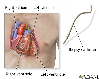
When a small piece of heart muscle tissue is needed for examination, a heart biopsy can be performed. A catheter is carefully threaded into an artery or vein to gain access into the heart. A bioptome (catheter with jaws in its tip) is then introduced. Once the bioptome is in place, three to five small pieces of tissue from the heart muscle are removed. The test is performed routinely after heart transplantation to detect potential rejection. It may also be performed when cardiomyopathy, myocarditis, cardiac amyloidosis, or other disorders are suspected.
Illustration
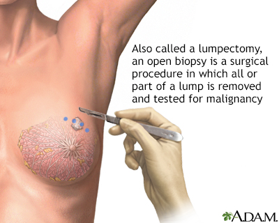
Open biopsy of the breast
An open biopsy can be performed under local or general anesthesia and will leave a small scar. Prior to surgery, a radiologist often first marks the lump with a wire, making it easier for the surgeon to find.
Illustration
Needle biopsy of the breast
A needle biopsy is performed under local anesthesia. Simple aspirations are performed with a small gauge needle to attempt to draw fluid from lumps that are thought to be cysts. Fine needle biopsy uses a larger needle to make multiple passes through a lump, drawing out tissue and fluid. Withdrawn fluid and tissue is further evaluated to determine if there are cancerous cells present.
Illustration
Core needle biopsy of the breast
A core needle biopsy of the breast is a procedure to remove samples of tissue from a lump or suspicious area of the breast and evaluate it for breast cancer. Core needle biopsy uses a long, hollow needle to take several core samples of tissue, usually using ultrasound or mammographic guidance. The samples are then sent to a lab for analysis.
Illustration
Testicular biopsy
Testicular biopsy is a procedure in which a small portion of testicle is removed for examination. The biopsy is performed by creating a small incision in the skin of the scrotum. A small piece of the testicle tissue is removed through the incision by snipping the sample off with small scissors. The test is usually performed when a semen analysis suggests that there is abnormal sperm, and other tests have not determined the cause. It may also be performed when testicular self-examination has revealed a lump.
Illustration
Rectal biopsy
Rectal biopsy can be used to determine the cause of blood, mucus, or pus in the stool. Rectal biopsy can also confirm findings of another test or x-rays, or take a biopsy of a growth found in the colon.
Illustration
Incision for pleural tissue biopsy
In an open pleural biopsy, a small piece of the pleural tissue is removed through a surgical incision in the chest. After the sample is obtained, a chest tube is placed and the incision is closed with stitches. Abnormal results may indicate tuberculosis, abnormal growths, viral, fungal, and parasitic diseases.
Illustration
Mucosal biopsy
Mucosal skin biopsy is the removal of a small piece of skin or mucous membrane. The sample can be retrieved in several ways: a shave biopsy (scraping or shaving a thin layer), a punch biopsy (using a needle or punch to obtain a small, but deeper, sample), or an excision of tissue (cutting to remove a piece of tissue). The sample is sent to the laboratory to isolate and identify organisms that cause infection.
Illustration
Bone biopsy
A bone biopsy is performed by making a small incision into the skin. A biopsy needle retrieves a sample of bone and it is sent for examination. The most common reasons for bone lesion biopsy are to distinguish between benign and malignant bone tumors, and to identify other bone abnormalities. Bone biopsy may also be performed to determine the cause of bone pain and tenderness.
Illustration
Nasal biopsy
A nasal biopsy is a diagnostic procedure in which a small piece of tissue is removed from the mucosal lining of the nose. The biopsy is most often performed when abnormal tissue is observed during an examination of the nose, or when disorders affecting the nasal mucosal tissue are suspected.
Illustration
Review Date:
9/30/2024+
Reviewed By:
Jonas DeMuro, MD, Diplomate of the American Board of Surgery with added Qualifications in Surgical Critical Care, Assistant Professor of Surgery, Renaissance School of Medicine, Stony Brook, NY. Review provided by VeriMed Healthcare Network. Also reviewed by David C. Dugdale, MD, Medical Director, Brenda Conaway, Editorial Director, and the A.D.A.M. Editorial team.


 All rights reserved.