
MRI
Magnetic resonance imaging; Nuclear magnetic resonance (NMR) imagingA magnetic resonance imaging (MRI) scan is an imaging test that uses powerful magnets and radio waves to create pictures of the body. It does not use ionizing radiation (x-rays). Single MRI images are called slices. The images can be stored on a computer, viewed on a monitor, printed on film or scanned to a disk. One exam can produce thousands...
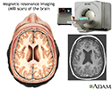
MRI of the brain
An MRI (magnetic resonance imaging) of the brain creates a detailed image of the complex structures in the brain. An MRI can give a three-dimensional depiction of the brain, making location of problems such as tumors or aneurysms more precise.
MRI of the brain
illustration
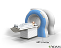
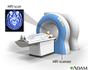
MRI scans
MRI stands for magnetic resonance imaging. It allows imaging of the interior of the body without using x-rays or other types of ionizing radiation. An MRI scan is capable of showing fine detail of different tissues.
MRI scans
illustration
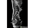
Teratoma - MRI scan
This MRI scan shows a tumor (teratoma) at the base of the spine (seen on the left lower edge of the screen), located in the sacrum and coccyx (sacrococcygeal) area. Teratomas are present at birth and may contain hair, teeth, and other tissues.
Teratoma - MRI scan
illustration
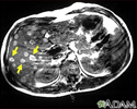
Melanoma of the liver - MRI scan
This upper abdominal MRI scan shows multiple tumors in the liver which have spread (metastasized) from a malignant melanoma in an arm or leg. Note the light circular areas throughout the liver (seen mostly on the left side of the screen).
Melanoma of the liver - MRI scan
illustration
Head MRI
MRI stands for magnetic resonance imaging. It allows imaging of the interior of the body without using x-rays or other types of ionizing radiation. An MRI scan is capable of showing fine detail of different tissues of the head.
Head MRI
illustration
MRI of the brain
An MRI (magnetic resonance imaging) of the brain creates a detailed image of the complex structures in the brain. An MRI can give a three-dimensional depiction of the brain, making location of problems such as tumors or aneurysms more precise.
MRI of the brain
illustration
MRI scans
MRI stands for magnetic resonance imaging. It allows imaging of the interior of the body without using x-rays or other types of ionizing radiation. An MRI scan is capable of showing fine detail of different tissues.
MRI scans
illustration
Teratoma - MRI scan
This MRI scan shows a tumor (teratoma) at the base of the spine (seen on the left lower edge of the screen), located in the sacrum and coccyx (sacrococcygeal) area. Teratomas are present at birth and may contain hair, teeth, and other tissues.
Teratoma - MRI scan
illustration
Melanoma of the liver - MRI scan
This upper abdominal MRI scan shows multiple tumors in the liver which have spread (metastasized) from a malignant melanoma in an arm or leg. Note the light circular areas throughout the liver (seen mostly on the left side of the screen).
Melanoma of the liver - MRI scan
illustration
Head MRI
MRI stands for magnetic resonance imaging. It allows imaging of the interior of the body without using x-rays or other types of ionizing radiation. An MRI scan is capable of showing fine detail of different tissues of the head.
Head MRI
illustration
MRI
Magnetic resonance imaging; Nuclear magnetic resonance (NMR) imagingA magnetic resonance imaging (MRI) scan is an imaging test that uses powerful magnets and radio waves to create pictures of the body. It does not use ionizing radiation (x-rays). Single MRI images are called slices. The images can be stored on a computer, viewed on a monitor, printed on film or scanned to a disk. One exam can produce thousands...
Review Date: 7/15/2024
Reviewed By: Jason Levy, MD, FSIR, Northside Radiology Associates, Atlanta, GA. Also reviewed by David C. Dugdale, MD, Medical Director, Brenda Conaway, Editorial Director, and the A.D.A.M. Editorial team.
 All rights reserved.