Transvaginal ultrasound
Endovaginal ultrasound; Ultrasound - transvaginal; Fibroids - transvaginal ultrasound; Vaginal bleeding - transvaginal ultrasound; Uterine bleeding - transvaginal ultrasound; Menstrual bleeding - transvaginal ultrasound; Infertility - transvaginal ultrasound; Ovarian - transvaginal ultrasound; Abscess - transvaginal ultrasoundTransvaginal ultrasound is a test used to look at a woman's uterus, ovaries, tubes, cervix, and pelvic area.
Cervix
The cervix is the lower end of the womb (uterus). It is at the top of the vagina. It is about 2. 5 to 3. 5 centimeters (1 to 1. 3 inches) long. Th...

Transvaginal means across or through the vagina. The ultrasound probe will be placed inside the vagina during the test.
How the Test is Performed
You will lie down on your back on a table with your knees bent. Your feet may be held in stirrups.
The ultrasound technician or doctor will introduce an ultrasound probe into your vagina. It may be mildly uncomfortable, but will not hurt. The probe is covered with a condom and a gel.
- The probe transmits sound waves and records the reflections of those waves off body structures. The ultrasound machine creates an image of the body part.
- The image is displayed on the ultrasound machine. In many offices, the person having the test can see the image also.
- The health care provider will gently move the probe around the area to see the pelvic organs.
In some cases, a special transvaginal ultrasound method called saline infusion sonography (SIS) may be needed to more clearly view the uterus.
How to Prepare for the Test
You will be asked to undress, usually from the waist down. A transvaginal ultrasound is done with your bladder empty or partly filled.
How the Test will Feel
In most cases, there is no pain. Some women may have mild discomfort from the pressure of the probe. Only a small part of the probe is placed into the vagina.
Why the Test is Performed
Transvaginal ultrasound may be done for the following problems:
- Abnormal findings on a physical exam, such as cysts, fibroid tumors, or other growths
Fibroid
Uterine fibroids are tumors that grow in a woman's womb (uterus). These growths are typically not cancerous (benign), and do not become cancerous....
 ImageRead Article Now Book Mark Article
 - Abnormal vaginal bleeding and menstrual problems
- Certain types of infertility
Infertility
Infertility means you cannot get pregnant (conceive). There are 2 types of infertility:Primary infertility refers to couples who have not become preg...
 ImageRead Article Now Book Mark Article
 -
Ectopic pregnancy
Ectopic pregnancy
An ectopic pregnancy is a pregnancy that occurs outside the womb (uterus).
 ImageRead Article Now Book Mark Article
 - Pelvic pain
This ultrasound is also used during pregnancy.
This ultrasound is also used during pre...
A pregnancy ultrasound is an imaging test that uses sound waves to create a picture of how a baby is developing in the womb (uterus). It is also use...

Normal Results
The pelvic structures or fetus is normal.
What Abnormal Results Mean
An abnormal result may be due to many conditions. Some problems that may be seen include:
- Birth defects in an unborn baby
- Cancers of the uterus, ovaries, vagina, and other pelvic structures
- Infection, including pelvic inflammatory disease
- Benign growths in or around the uterus and ovaries (such as cysts or fibroids)
- Endometriosis
- Pregnancy outside of the uterus (ectopic pregnancy)
- Twisting of the ovaries
Risks
There are no known harmful effects of transvaginal ultrasound on humans.
Unlike traditional x-rays, there is no radiation exposure with this test.
References
Dolan MS, Hill CC, Valea FA. Benign gynecologic lesions: vulva, vagina, cervix, uterus, oviduct, ovary, ultrasound imaging of pelvic structures. In: Gershenson DM, Lentz GM, Valea FA, Lobo RA, eds. Comprehensive Gynecology. 8th ed. Philadelphia, PA: Elsevier; 2022:chap 18.
Hur HC, Lobo RA. Ectopic pregnancy: etiology, pathology, diagnosis, management, fertility prognosis. In: Gershenson DM, Lentz GM, Valea FA, Lobo RA, eds. Comprehensive Gynecology. 8th ed. Philadelphia, PA: Elsevier; 2022:chap 17.
Kelly CM. Ectopic pregnancy. In: Kellerman RD, Rakel DP, Heidelbaugh JJ, Lee EM, eds. Conn's Current Therapy 2024. Philadelphia, PA: Elsevier; 2024:1288-1291.
Wei PK, Savicke AM, Levine D. The uterus. In: Rumack CM, Levine D, eds. Diagnostic Ultrasound. 6th ed. Philadelphia, PA: Elsevier; 2024:chap 28.
-
Ultrasound in pregnancy - illustration
The ultrasound has become a standard procedure used during pregnancy. It can demonstrate fetal growth and can detect increasing numbers of conditions including meningomyelocele, congenital heart disease, kidney abnormalities, hydrocephalus, anencephaly, club feet, and other deformities. Ultrasound does not produce ionizing radiation and is considered a very safe procedure for both the mother and the fetus.
Ultrasound in pregnancy
illustration
-
Female reproductive anatomy - illustration
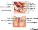
Internal structures of the female reproductive anatomy include the uterus, ovaries, and cervix. External structures include the labium minora and majora, the vagina and the clitoris.
Female reproductive anatomy
illustration
-
Uterus - illustration
The uterus is a hollow muscular organ located in the female pelvis between the bladder and rectum. The ovaries produce the eggs that travel through the fallopian tubes. Once the egg has left the ovary it can be fertilized and implant itself in the lining of the uterus. The main function of the uterus is to nourish the developing fetus prior to birth.
Uterus
illustration
-
Transvaginal ultrasound - illustration
Transvaginal ultrasound is a method of imaging the genital tract in females. A hand held probe is inserted directly into the vagina. The probe is moved within the vaginal cavity to scan the pelvic structures, while ultrasound pictures are viewed on a monitor. The test can be performed to evaluate women with infertility problems, abnormal bleeding, sources of unexplained pain, congenital malformations of the uterus and ovaries, and possible tumors and infection.
Transvaginal ultrasound
illustration
-
Ultrasound in pregnancy - illustration
The ultrasound has become a standard procedure used during pregnancy. It can demonstrate fetal growth and can detect increasing numbers of conditions including meningomyelocele, congenital heart disease, kidney abnormalities, hydrocephalus, anencephaly, club feet, and other deformities. Ultrasound does not produce ionizing radiation and is considered a very safe procedure for both the mother and the fetus.
Ultrasound in pregnancy
illustration
-
Female reproductive anatomy - illustration
Internal structures of the female reproductive anatomy include the uterus, ovaries, and cervix. External structures include the labium minora and majora, the vagina and the clitoris.
Female reproductive anatomy
illustration
-
Uterus - illustration
The uterus is a hollow muscular organ located in the female pelvis between the bladder and rectum. The ovaries produce the eggs that travel through the fallopian tubes. Once the egg has left the ovary it can be fertilized and implant itself in the lining of the uterus. The main function of the uterus is to nourish the developing fetus prior to birth.
Uterus
illustration
-
Transvaginal ultrasound - illustration
Transvaginal ultrasound is a method of imaging the genital tract in females. A hand held probe is inserted directly into the vagina. The probe is moved within the vaginal cavity to scan the pelvic structures, while ultrasound pictures are viewed on a monitor. The test can be performed to evaluate women with infertility problems, abnormal bleeding, sources of unexplained pain, congenital malformations of the uterus and ovaries, and possible tumors and infection.
Transvaginal ultrasound
illustration
Review Date: 4/16/2024
Reviewed By: John D. Jacobson, MD, Professor Emeritus, Department of Obstetrics and Gynecology, Loma Linda University School of Medicine, Loma Linda, CA. Also reviewed by David C. Dugdale, MD, Medical Director, Brenda Conaway, Editorial Director, and the A.D.A.M. Editorial team.


