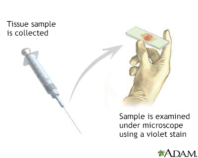
Gram stain of tissue biopsy
Tissue biopsy - Gram stain
Gram stain of tissue biopsy test involves using crystal violet stain to test a sample of tissue taken from a biopsy.
The Gram stain method can be used on almost any specimen. It is an excellent technique for making a general, basic identification of the type of bacteria in the sample.
Images

I Would Like to Learn About:
How the Test is Performed
A sample, called a smear, from a tissue specimen is placed in a very thin layer on a microscope slide. The specimen is stained with crystal violet stain and goes through more processing before it is examined under the microscope for bacteria.
The characteristic appearance of the bacteria, such as their color, shape, clustering (if any), and pattern of staining help determine the type of bacteria.
How to Prepare for the Test
If the biopsy is included as part of a surgical procedure, you will be asked not to eat or drink anything the night before surgery. If the biopsy is of a superficial (on the surface of the body) tissue, you may be asked not to eat or drink for several hours before the procedure.
How the Test will Feel
How the test feels depends on the part of the body being biopsied. There are several different methods for taking tissue samples.
- A needle may be inserted through the skin to the tissue.
- A cut (incision) through the skin into the tissue may be made, and a small piece of the tissue removed.
- A biopsy may also be taken from inside the body using an instrument that helps the doctor see inside the body, such as an endoscope or cystoscope.
You may feel pressure and mild pain during a biopsy. Some form of pain-relieving medicine (anesthetic) is usually given, so you have little or no pain.
Why the Test is Performed
The test is performed when an infection of a body tissue is suspected.
Normal Results
Whether there are bacteria, and what type there are, depends on the tissue being biopsied. Some tissues in the body are sterile, such as the brain. Other tissues, such as the gut, normally contain bacteria.
Note: Normal value ranges may vary slightly among different laboratories. Talk to your doctor about the meaning of your specific test results.
What Abnormal Results Mean
Abnormal results usually mean there is an infection in the tissue. More tests, such as culturing the tissue that was removed, are often needed to identify the type of bacteria.
Risks
Risks depend on the procedure used to take the tissue biopsy, and may include bleeding or infection.
Related Information
BiopsyReferences
Chernecky CC, Berger BJ. Biopsy, site-specific - specimen. In: Chernecky CC, Berger BJ, eds. Laboratory Tests and Diagnostic Procedures. 6th ed. St Louis, MO: Elsevier Saunders; 2013:199-202.
Wojewoda CM, Stempak LM. Medical bacteriology. In: McPherson RA, Pincus MR, eds. Henry's Clinical Diagnosis and Management by Laboratory Methods. 24th ed. Philadelphia, PA: Elsevier; 2022:chap 57.
BACK TO TOPReview Date: 12/4/2022
Reviewed By: Jatin M. Vyas, MD, PhD, Associate Professor in Medicine, Harvard Medical School; Associate in Medicine, Division of Infectious Disease, Department of Medicine, Massachusetts General Hospital, Boston, MA. Also reviewed by David C. Dugdale, MD, Medical Director, Brenda Conaway, Editorial Director, and the A.D.A.M. Editorial team.

Health Content Provider
06/01/2025
|
A.D.A.M., Inc. is accredited by URAC, for Health Content Provider (www.urac.org). URAC's accreditation program is an independent audit to verify that A.D.A.M. follows rigorous standards of quality and accountability. A.D.A.M. is among the first to achieve this important distinction for online health information and services. Learn more about A.D.A.M.'s editorial policy, editorial process and privacy policy. A.D.A.M. is also a founding member of Hi-Ethics. This site complied with the HONcode standard for trustworthy health information from 1995 to 2022, after which HON (Health On the Net, a not-for-profit organization that promoted transparent and reliable health information online) was discontinued. |
The information provided herein should not be used during any medical emergency or for the diagnosis or treatment of any medical condition. A licensed medical professional should be consulted for diagnosis and treatment of any and all medical conditions. Links to other sites are provided for information only -- they do not constitute endorsements of those other sites. © 1997- 2024 A.D.A.M., a business unit of Ebix, Inc. Any duplication or distribution of the information contained herein is strictly prohibited.
