
Vaginal cancer
Vaginal cancer; Cancer - vagina; Tumor - vaginalVaginal cancer is cancer of the vagina, a female reproductive organ.
Causes
Most vaginal cancers occur when another cancer, such as cervical, vulvar, or endometrial cancer, spreads. This is called secondary vaginal cancer.
Cervical
Cervical cancer is cancer that starts in the cervix. The cervix is the lower part of the uterus (womb) that opens at the top of the vagina.

Endometrial cancer
Endometrial cancer is cancer that starts in the endometrium, the lining of the uterus (womb).

Cancer that starts in the vagina is called primary vaginal cancer. This type of cancer is rare. Most primary vaginal cancers start in skin-like cells called squamous cells. This cancer is known as squamous cell carcinoma. The other types include:
- Adenocarcinoma
- Melanoma
- Sarcoma
The cause of squamous cell carcinoma of the vagina is unknown. But a history of cervical cancer is common in women with squamous cell carcinoma of the vagina. So it may be associated with human papilloma virus (HPV) infection.
Most women with squamous cell cancer of the vagina are over 50.
Adenocarcinoma of the vagina tends to affect younger women. The average age at which this cancer is diagnosed is 19. Women whose mothers took the medicine diethylstilbestrol (DES) to prevent miscarriages during the first 3 months of pregnancy are more likely to develop vaginal adenocarcinoma.
Miscarriages
A miscarriage is the spontaneous loss of a fetus before the 20th week of pregnancy. Pregnancy losses after the 20th week are called stillbirths. Mi...

Sarcoma of the vagina is a rare cancer that mainly occurs in infancy and early childhood.
Symptoms
Symptoms of vaginal cancer can include any of the following:
- Bleeding after having sex
- Painless vaginal bleeding and discharge not due to normal period
- Pain in the pelvis or vagina
Some women have no symptoms.
Exams and Tests
In women with no symptoms, the cancer may be found during a routine pelvic exam and Pap test.
Pap test
The Pap test mainly checks for changes that may turn into cervical cancer. Cells scraped from the opening of the cervix are examined under a microsc...

Other tests to diagnose vaginal cancer include:
-
Biopsy
Biopsy
A biopsy is the removal of a small piece of tissue for laboratory examination.
Read Article Now Book Mark Article -
Colposcopy
Colposcopy
A colposcopy is a special way of looking at the cervix. It uses a light and a low-powered microscope to make the cervix appear much larger. This he...
ImageRead Article Now Book Mark Article
Other tests that may be done to check if the cancer has spread include:
-
Chest x-ray
Chest x-ray
A chest x-ray is an x-ray of the chest, lungs, heart, large arteries, ribs, and diaphragm.
 ImageRead Article Now Book Mark Article
 -
CT scan and MRI of the abdomen and pelvis
CT scan
A computed tomography (CT) scan is an imaging method that uses x-rays to create pictures of cross-sections of the body. Related tests include:Abdomin...
 ImageRead Article Now Book Mark Article
MRI
A magnetic resonance imaging (MRI) scan is an imaging test that uses powerful magnets and radio waves to create pictures of the body. It does not us...
 ImageRead Article Now Book Mark Article
 -
PET scan
PET scan
A positron emission tomography (PET) scan is a type of imaging test. It uses a radioactive substance called a tracer to look for disease in the body...
Read Article Now Book Mark Article
Other tests that may be done to know the stage of the vaginal cancer include:
-
Cystoscopy
Cystoscopy
Cystoscopy is a surgical procedure. This is done to see the inside of the bladder and urethra using a thin, lighted tube.
 ImageRead Article Now Book Mark Article
 -
Barium enema
Barium enema
Barium enema is a special x-ray of the large intestine, which includes the colon and rectum.
 ImageRead Article Now Book Mark Article
 -
Intravenous urography (x-ray of kidney, ureters and bladder using contrast material)
Intravenous urography
An intravenous pyelogram (IVP) is a special x-ray exam of the kidneys, bladder, and ureters (the tubes that carry urine from the kidneys to the bladd...
 ImageRead Article Now Book Mark Article
Treatment
Treatment of vaginal cancer depends on:
- The type of cancer
- How far the disease has spread
Surgery is sometimes used if the cancer is small and located at the upper part of the vagina. Most women are treated with radiation. If the tumor is cervical cancer that has spread to the vagina, radiation and chemotherapy are both given.
Radiation
Radiation therapy uses high-powered radiation (such as x-rays or gamma rays), particles, or radioactive seeds to kill cancer cells.

Chemotherapy
The term chemotherapy is used to describe cancer-killing drugs. Chemotherapy may be used to:Cure the cancerShrink the cancerPrevent the cancer from ...

Sarcoma may be treated with a combination of chemotherapy, surgery, and radiation.
Support Groups
You can ease the stress of illness by joining a support group whose members share common experiences and problems.
Support group
The following organizations are good resources for information on cancer:American Cancer Society -- www. cancer. orgAmerican Childhood Cancer Organiz...
Read Article Now Book Mark ArticleOutlook (Prognosis)
The outlook for women with vaginal cancer depends on the size and the stage of disease and the specific type of tumor.
Possible Complications
Vaginal cancer may spread to other areas of the body. Complications can occur from radiation, surgery, and chemotherapy.
When to Contact a Medical Professional
Call for an appointment with your health care provider if:
- You notice bleeding after sex
- You have persistent vaginal bleeding or discharge
Prevention
There are no definite ways to prevent this cancer.
The HPV vaccine is approved to help prevent cervical cancer. This vaccine may also decrease the risk of getting some other HPV-associated cancers, such as vaginal cancer. You can increase your chance of early detection by getting regular pelvic examinations and Pap smears.
HPV vaccine
All content below is taken in its entirety from the CDC HPV (Human Papillomavirus) Vaccine Information Statement (VIS): www. cdc. gov/vaccines/hcp/vi...

References
Bodurka DC, Frumovitz M. Malignant diseases of the vagina: intraepithelial neoplasia, carcinoma, sarcoma. In: Lobo RA, Gershenson DM, Lentz GM, Valea FA, eds. Comprehensive Gynecology. 7th ed. Philadelphia, PA: Elsevier; 2017:chap 31.
Jhingran A, Russell AH, Seiden MV, et al. Cancers of the cervix, vulva, and vagina. In: Niederhuber JE, Armitage JO, Kastan MB, Doroshow JH, Tepper JE, eds. Abeloff's Clinical Oncology. 6th ed. Philadelphia, PA: Elsevier; 2020:chap 84.
PDQ Adult Treatment Editorial Board. Vaginal Cancer Treatment (PDQ®): Health Professional Version. 2022 Feb 24. In: PDQ Cancer Information Summaries [Internet]. Bethesda (MD): National Cancer Institute (US); 2002–. PMID: 26389242 pubmed.ncbi.nlm.nih.gov/26389242/.
-
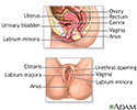
Female reproductive anatomy - illustration
Internal structures of the female reproductive anatomy include the uterus, ovaries, and cervix. External structures include the labium minora and majora, the vagina and the clitoris.
Female reproductive anatomy
illustration
-
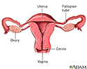
Uterus - illustration
The uterus is a hollow muscular organ located in the female pelvis between the bladder and rectum. The ovaries produce the eggs that travel through the fallopian tubes. Once the egg has left the ovary it can be fertilized and implant itself in the lining of the uterus. The main function of the uterus is to nourish the developing fetus prior to birth.
Uterus
illustration
-
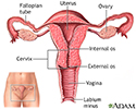
Normal uterine anatomy (cut section) - illustration
The uterus is a muscular organ with thick walls, two upper openings to the fallopian tubes and an inferior opening to the vagina.
Normal uterine anatomy (cut section)
illustration
-
Female reproductive anatomy - illustration
Internal structures of the female reproductive anatomy include the uterus, ovaries, and cervix. External structures include the labium minora and majora, the vagina and the clitoris.
Female reproductive anatomy
illustration
-
Uterus - illustration
The uterus is a hollow muscular organ located in the female pelvis between the bladder and rectum. The ovaries produce the eggs that travel through the fallopian tubes. Once the egg has left the ovary it can be fertilized and implant itself in the lining of the uterus. The main function of the uterus is to nourish the developing fetus prior to birth.
Uterus
illustration
-
Normal uterine anatomy (cut section) - illustration
The uterus is a muscular organ with thick walls, two upper openings to the fallopian tubes and an inferior opening to the vagina.
Normal uterine anatomy (cut section)
illustration
Review Date: 1/1/2022
Reviewed By: Howard Goodman, MD, Gynecologic Oncology, Florida Cancer Specialists & Research Institute, West Palm Beach, FL. Review provided by VeriMed Healthcare Network. Also reviewed by David Zieve, MD, MHA, Medical Director, Brenda Conaway, Editorial Director, and the A.D.A.M. Editorial team.
 All rights reserved.