Multimedia Gallery
Heart formation
The embryo’s heart is the first organ that forms in its tiny body, and like most complex instruments, it begins with some simple structures.
Let’s go back to 18 days after conception...Looking in the mother’s uterus, you can see the embryo surrounded by its yolk sac and amnion. Let’s take a look inside.
Here’s a diagram of the embryo seen from a side view. Right now, it’s about the size of a raisin. There’s the head region and that red-colored area slightly above it contains two tubes that will form the embryo’s heart. Here’s what the tubes look like from a front view.
On day 21, we see that the primitive heart tubes have moved below the embryo’s developing head region. And by day 22, the tubes have fused together, and have moved to the area that will eventually be our embryo’s thoracic, or chest cavity. It’s also about this time that the heart begins to beat for the first time...
Here’s what it looks like from the front.
Now let’s go back to day 18 and watch this happen from a different viewpoint. Here are two tubes in our embryo’s chest region seen from a front view. Watch this... Over the next two days, these tubes fuse together.
Here’s another amazing part: the tube now starts bending and twisting and over the next 8 days it forms a simple version of the heart.
By the time the embryo becomes a fetus at two months, the heart bears a close resemblance to what it will look like after the baby’s born. But the resemblance is only superficial. On the inside of the heart, things are much different in both form and function.
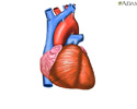
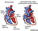
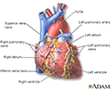
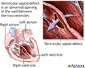
Here’s a newborn heart on the left. Let’s take a closer look. There’s the right atrium right ventricle, left atrium and left ventricle. The two major blood vessels are the aorta and the pulmonary artery.
The pathway of blood in the newborn heart works like this: oxygen-poor blood from the body enters the right atrium, then goes to the right ventricle. From the right ventricle, the blood is pumped to the lungs where it becomes oxygen rich. Then the blood flows back to the heart filling the left atrium and from there on to the left ventricle. The left ventricle pumps the oxygen rich blood through the aorta, which carries it to the rest of the newborn’s body.
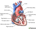
You can see the fetal heart has the same basic components as the newborn heart, but there are a couple important differences. Because the placenta is providing all of the oxygen the fetus requires, its lungs are not needed to perform this task, and therefore much of the fetus’ blood is detoured away from the lungs through two openings or connections. They are the foramen ovale, which connects the right and left atria, and the ductus arteriosus which connects the aorta and the pulmonary artery.
As blood enters the heart into the right atrium some of the blood flows into the right ventricle as in the newborn, but also notice that some blood flows directly into the left atrium through the foramen ovale. This blood will pass directly into the left ventricle and be pumped out to the body without ever having gone to the lungs. In addition, some of the blood that did enter the right ventricle, and would normally go to the lungs, never reaches the lungs.
Here lets watch. As blood is being pumped out of the right ventricle towards the lungs through the pulmonary artery, some of that blood escapes into the aorta through the ductus arteriosus, bypassing the lungs as it does. These two important connections will remain open up until the time of birth.
Within thirty minutes after the baby’s first breath, the ductus arteriosus will completely close, and the flap of the foramen ovale will shut off like a valve. This happens because of an increase in pressure on the left side of the heart, and a decrease on the right side. These changes in the heart anatomy cause the blood to flow to the lungs, which will take over their lifelong job of supplying oxygen to the body.
It’s incredible to think that this complex organ started off as a couple of tubes only 2 1/2 weeks ago.
Heart formation
Review Date: 11/2/2021
Reviewed By: Thomas S. Metkus, MD, Assistant Professor of Medicine and Surgery, Johns Hopkins University School of Medicine, Baltimore, MD. Also reviewed by David Zieve, MD, MHA, Medical Director, Brenda Conaway, Editorial Director, and the A.D.A.M. Editorial team.
Animations
- Abdominal aortic aneurysm
- Abdominal pain
- Aneurysm description
- Arrhythmias
- Atherosclerosis
- Atrial fibrillation
- Balloon angioplasty - short...
- Blood clotting
- Blood flow
- Blood pressure
- Brain components
- Cardiac and vascular disord...
- Cardiac arrhythmia - conduc...
- Cardiac arrhythmia symptoms
- Cardiac arrhythmia tests: E...
- Cardiac arrhythmia: Additio...
- Cardiac arrhythmia: Heart p...
- Cardiac arrhythmia: Physica...
- Cardiac arrhythmia: Taking ...
- Cardiac catheterization
- Cardiac catheterization - a...
- Cardiac conduction system
- Cardiac CT scan overview
- Cardiomyopathy
- Cardiovascular system
- Causes and side effects of ...
- Cerebral aneurysm
- Chest pain
- Childhood obesity
- Cholesterol and triglycerid...
- Coronary artery bypass graf...
- Coronary artery disease
- Coronary artery disease (CA...
- Directional coronary athere...
- Electrocardiogram
- Epinephrine and exercise
- Erection problems
- Essential hypertension
- Exercise
- Hardening of arteries
- Healthy Guide to Fast Food
- Heart attack
- Heart bypass surgery
- Heart failure
- Heart formation
- Heartbeat
- How to use a pill cutter
- Hypertension
- Hypertension - overview
- Muscle types
- NICU consultants and suppor...
- Nuclear stress test
- Obstructive sleep apnea
- Percutaneous transluminal c...
- Preeclampsia
- Smoking
- Smoking tips to quit
- Snoring
- Stent
- Stroke
- Stroke
- Stroke - secondary to cardi...
- Tachycardia
- Tobacco use - effects on ar...
- Tracking your blood pressur...
- Type 2 diabetes
- Understanding cholesterol r...
- Vacation health care
- Valvular heart disease (VHD...
- Varicose veins
- Varicose veins overview
- Venous insufficiency
- What makes your heart beat?
Illustrations
- 15/15 rule
- Absent pulmonary valve
- Acute MI
- Adjustable gastric banding
- Aerobic exercise
- Alcoholic cardiomyopathy
- Alpha-glucosidase inhibitors
- Angina
- Anomalous left coronary artery
- Anterior heart arteries
- Aortic aneurysm
- Aortic dissection
- Aortic rupture - chest x-ray
- Aortic stenosis
- Aortopulmonary window
- Arterial embolism
- Arterial plaque build-up
- Arterial tear in internal c...
- Arteries of the brain
- Artery cut section
- Atherosclerosis
- Atherosclerosis of internal...
- Atherosclerosis of the extr...
- Atrial septal defect
- Atrioventricular block - EC...
- Atrioventricular canal (end...
- Auscultation
- Bacterial pericarditis
- Balance receptors
- Bicuspid aortic valve
- Biguanides
- Biliopancreatic diversion (BPD)
- Biliopancreatic diversion w...
- Biopsy catheter
- Blood pressure
- Blood pressure check
- Blood test
- Bradycardia
- Brain
- Brainstem function
- Breathing
- Bronchial cancer - CT scan
- Calcium benefit
- Calcium source
- Calories and fat per serving
- Cardiac arteriogram
- Cardiac catheterization
- Carotid dissection
- Carotid duplex
- Carotid stenosis - X-ray of...
- Cataract
- Cataract - close-up of the eye
- Central nervous system and ...
- Cerebellum - function
- Cerebral aneurysm
- Cholesterol
- Cholesterol producers
- Circle of Willis
- Circulation of blood throug...
- Circulatory system
- Clubbing
- Coarctation of the aorta
- Conduction system of the heart
- Constrictive pericarditis
- Coronary angiography
- Coronary artery blockage
- Coronary artery disease
- Coronary artery fistula
- Coronary artery spasm
- Coronary artery stent
- Crossed eyes
- CT scan
- Culture-negative endocarditis
- Cyanosis of the nail bed
- Cyanotic heart disease
- DASH diet
- Deep veins
- Deep veins
- Deep venous thrombosis - il...
- Developmental process of at...
- Dextrocardia
- Diabetes and exercise
- Diabetic emergency supplies
- Digestive system
- Dilated cardiomyopathy
- Double aortic arch
- Double inlet left ventricle
- Double outlet right ventricle
- Drug induced hypertension
- Duplex/doppler ultrasound test
- Ear anatomy
- Ebstein's anomaly
- ECG
- ECMO
- Effects of age on blood pressure
- Eisenmenger syndrome (or co...
- Electrocardiogram (ECG)
- Emphysema
- Endarterectomy
- Endocrine glands
- Enlarged view of atherosclerosis
- Exercise - a powerful tool
- Exercise 30 minutes a day
- Exercise can lower blood pr...
- Exercise with friends
- Eye
- Facial drooping
- Fast food
- Fish in diet
- Food and insulin release
- Food label guide for candy
- Food label guide for whole ...
- Foot swelling
- Fruits and vegetables
- Glucose in blood
- Glucose test
- Healthy diet
- Healthy diet
- Heart - front view
- Heart - section through the...
- Heart attack symptoms
- Heart beat
- Heart chambers
- Heart valves
- Heart valves - anterior view
- Heart valves - superior view
- High blood pressure tests
- Holter heart monitor
- Hypertension
- Hypertensive kidney
- Hypertrophic cardiomyopathy
- Infective endocarditis
- Insulin pump
- Insulin pump
- Intracerebellar hemorrhage ...
- Intracerebral hemorrhage
- Janeway lesion on the finger
- Jaw pain and heart attacks
- Left atrial myxoma
- Left cerebral hemisphere - ...
- Left heart catheterization
- Leg pain (Osgood-Schlatter)
- Lifestyle changes
- Lobes of the brain
- Low blood sugar symptoms
- Low nasal bridge
- Lower leg edema
- Lower leg muscles
- Lung mass, right lung - CT scan
- Lung mass, right upper lobe...
- Lung nodule, right lower lu...
- Lung with squamous cell can...
- Lungs
- Lymph tissue in the head an...
- Male reproductive anatomy
- Mitral stenosis
- Mitral valve prolapse
- Monitoring blood pressure
- MRI scans
- MUGA test
- Muscular atrophy
- myPlate
- Neck pain
- Neck pulse
- Normal anatomy of the heart
- Normal heart anatomy (cut s...
- Normal heart rhythm
- Normal lung anatomy
- Omega-3 fatty acids
- Otoscope examination
- Pacemaker
- Pericarditis
- Pericardium
- Pericardium
- Peripartum cardiomyopathy
- Pharmacy options
- Pitting edema on the leg
- Plaque buildup in arteries
- Post myocardial infarction ...
- Posterior heart arteries
- Post-MI pericarditis
- Prevention of heart disease
- Progressive build-up of pla...
- Ptosis - drooping of the eyelid
- Pulmonary nodule, solitary ...
- Quitting smoking
- Radial pulse
- Read food labels
- Respiratory cilia
- Respiratory system
- Retrocalcaneal bursitis
- Right atrial myxoma
- Right cerebral hemisphere -...
- Roux-en-Y stomach surgery f...
- Saturated fats
- Secondhand smoke and lung cancer
- Shin splints
- Slit-lamp exam
- Smoking hazards
- Smoking hazards
- Sodium content
- Sources of fiber
- Stable angina
- Starchy foods
- Striae in the popliteal fossa
- Stroke
- Sulfonylureas drug
- Superficial thrombophlebitis
- Swan Ganz catheterization
- Taking your carotid pulse
- Thiazolidinediones
- Thoracic organs
- Thromboangiitis obliterans
- Thrombus
- Thyroid cancer - CT scan
- Tobacco and cancer
- Tobacco and chemicals
- Tobacco and vascular disease
- Tobacco health risks
- Totally anomalous pulmonary...
- Trans fatty acids
- Transient Ischemic attack (TIA)
- Tricuspid Regurgitation
- Type I diabetes
- Ultrasound, normal fetus - ...
- Ultrasound, ventricular sep...
- Untreated hypertension
- Varicose veins
- Vascular ring
- Venous blood clot
- Ventricular septal defect
- Ventricular tachycardia
- Vertebra, thoracic (mid back)
- Vertical banded gastroplasty
- Vertigo
- Visual acuity test
- Visual field test
- Vitamin B1 benefit
- Vitamin B1 source
- Vitamin C benefit
- Vitamin C deficit
- Vitamin C source
- Vitamin E and heart disease
- Warming up and cooling down
- Wine and health