Spinal injury
Spinal cord injury; SCIThe spinal cord contains the nerves that carry messages between your brain and the rest of the body. The cord passes through your neck and back. A spinal cord injury is very serious because it can cause loss of movement (paralysis), function, and sensation below the site of the injury.
Spinal cord injury
Spinal cord trauma is damage to the spinal cord. It may result from direct injury to the cord itself or indirectly from disease of the nearby bones,...

Causes
A spinal cord injury may be caused by incidents such as:
- Bullet or stab wound
- Fracture of the spine
- Traumatic injury to the face, neck, head, chest, or back (for example, a car accident)
- Diving accident
- Electric shock
Electric shock
An electrical injury is damage to the skin or internal organs when a person comes into direct contact with an electrical current.
 ImageRead Article Now Book Mark Article
 - Extreme twisting of the middle of the body
- Sports injury
- Falls
Symptoms
Symptoms of a spinal cord injury may include any of the following:
- Head that is in an unusual position
- Numbness or tingling that spreads down an arm or leg
- Weakness
- Difficulty walking
- Paralysis (loss of movement) of arms or legs
Paralysis
Muscle function loss is when a muscle does not work or move normally. The medical term for complete loss of muscle function is paralysis.
 ImageRead Article Now Book Mark Article
 - Loss of bladder or bowel control
- Shock (pale, clammy skin, bluish lips and fingernails, acting dazed or semiconscious)
Shock
Shock is a life-threatening condition that occurs when the body is not getting enough blood flow. Lack of blood flow means the cells and organs do n...
 ImageRead Article Now Book Mark Article
 - Lack of alertness (unconsciousness)
- Stiff neck, headache, or neck pain
First Aid
Never move anyone who you think may have a spinal injury unless it is absolutely necessary. For example, if you need to get the person out of a burning car or help them to breathe.
Keep the person completely still and safe until medical help arrives.
- Call the local emergency number, such as 911.
- Hold the person's head and neck in the position in which they were found. DO NOT try to straighten the neck. DO NOT allow the neck to bend or twist.
- DO NOT allow the person to get up and walk.
If the person is not alert or responding to you:
- Check the person's breathing and circulation.
- If needed, do CPR. DO NOT do rescue breathing or change the position of the neck, do chest compressions only.
CPR
CPR stands for cardiopulmonary resuscitation. It is an emergency life-saving procedure that is done when someone's breathing or heartbeat has stoppe...
Read Article Now Book Mark Article
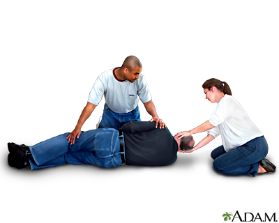
DO NOT roll the person over unless the person is vomiting or choking on blood, or you need to check for breathing.
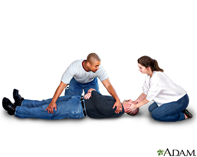
If you need to roll the person over:
- Have someone help you.so that you can turn the person without moving the neck and head relative to the body.
- One person should be located at the person's head, the other at the person's side.
- Keep the person's head, neck, and back in line while you roll them onto one side.
Do Not
- DO NOT bend, twist, or lift the person's head or body.
- DO NOT attempt to move the person before medical help arrives unless it is absolutely necessary.
- DO NOT remove a football helmet or pads if a spinal injury is suspected.
When to Contact a Medical Professional
Call your local emergency number (such as 911) if you think someone has a spinal cord injury. DO NOT move the person unless there is urgent danger.
Prevention
The following may lower your risk for spinal injury:
- Wear seat belts.
- Do not drink and drive.
- Do not dive into pools, lakes, rivers, and other bodies of water, particularly if you cannot determine the depth of the water or if the water is not clear.
- Do not tackle or dive into a person with your head.
References
National Institute of Neurological Disorders and Stroke website. Spinal cord injury. www.ninds.nih.gov/health-information/disorders/spinal-cord-injury. Last reviewed October 16, 2023. Accessed October 16, 2023.
Preston-Suni K, Kaji AH. Spinal trauma. In: Walls RM, ed. Rosen's Emergency Medicine: Concepts and Clinical Practice. 10th ed. Philadelphia, PA: Elsevier; 2023:chap 35.
Willians KD. Fractures, dislocations, and fracture-dislocations of the spine. In: Azar FM, Beaty JH, eds. Campbell's Operative Orthopaedics. 14th ed. Phildelphia, PA: Elsevier; 2021:chap 41.
Skeletal spine - illustration
The spine is divided into several sections. The cervical vertebrae make up the neck. The thoracic vertebrae comprise the chest section and have ribs attached. The lumbar vertebrae are the remaining vertebrae below the last thoracic bone and the top of the sacrum. The sacral vertebrae are caged within the bones of the pelvis, and the coccyx represents the terminal vertebrae or vestigial tail.
Skeletal spine
illustration
Vertebra, cervical (neck) - illustration
These are the seven bones of the neck, called the cervical vertebra. The top bone, seen on the right of this picture, is called the atlas, and is where the head attaches to the neck. The second bone is called the axis, upon which the head and atlas rotate. The vertebra are numbered from one to seven from the atlas down, and are referred to as C1, C2, C3, etc.
Vertebra, cervical (neck)
illustration
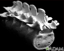
Vertebra, lumbar (low back) - illustration
These are the five vertebra of the lower back. The last vertebra (on the upper left of the picture) attaches to the sacrum, and the top vertebra (on the right of the picture) attaches to the thoracic section of the back. The vertebra are broader and stronger than the other bones in the spine. This allows them to absorb the added pressure applied to the lower back, but this area remains a common site of injury. The vertebra are numbered from one to five and are labeled L1, L2, L3 etc. from the higher bones to the lower.
Vertebra, lumbar (low back)
illustration
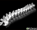
Vertebra, thoracic (mid back) - illustration
These are twelve vertebra of the mid back. The last vertebra (on the left side of the picture) attaches to the lumbar (lower) spine, and the top vertebra (on the right) attaches to the cervical (neck) section of the back. The vertebra are broader and stronger than the cervical bones. This allows them to absorb the added pressure applied to the mid back, but they remain a common sight of injury. The vertebra are numbered from one to twelve and labeled T1, T2, T3, et cetera, from the upper most bones to the lowest.
Vertebra, thoracic (mid back)
illustration
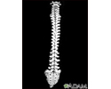
Vertebral column - illustration
This is the spine and the sacrum with the cervical (neck), thoracic (mid-back), and lumbar (lower back) vertebra. Notice how the appearance of the vertebra change as you look down the spine. The change in shape and size reflect the different functions of the neck, mid-back, and lower back.
Vertebral column
illustration
Central nervous system - illustration
The central nervous system is made up of the brain and spinal cord. The brain functions to receive nerve impulses from the spinal cord and cranial nerves. The spinal cord contains the nerves that carry messages between the brain and the body. Spinal cord injury can occur when there is damage to the cells within the spinal cord or when the tracts of nerves that run up and down the spinal cord are severed.
Central nervous system
illustration
Spinal cord injury - illustration
A severe spinal cord injury often causes loss of feeling and paralysis, the loss of movement and voluntary control over the muscles in the body. Spinal cord damage also causes loss of reflex function below the point of injury interrupting bodily functions such as breathing, bowel control, and bladder control. In the event of a spinal injury prompt medical attention can help to minimize further spinal cord damage.
Spinal cord injury
illustration
Spinal anatomy - illustration
The vertebral column provides structural support for the trunk and surrounds and protects the spinal cord. The vertebral column also provides attachment points for the muscles of the back and ribs. The vertebral disks serve as shock absorbers during activities such as walking, running, and jumping. They also allow the spine to flex and extend.
Spinal anatomy
illustration
Two person roll - Procedure, part 1
Presentation
Review Date: 4/24/2023
Reviewed By: C. Benjamin Ma, MD, Professor, Chief, Sports Medicine and Shoulder Service, UCSF Department of Orthopaedic Surgery, San Francisco, CA. Also reviewed by David C. Dugdale, MD, Medical Director, Brenda Conaway, Editorial Director, and the A.D.A.M. Editorial team. Editorial update 10/16/2023.


